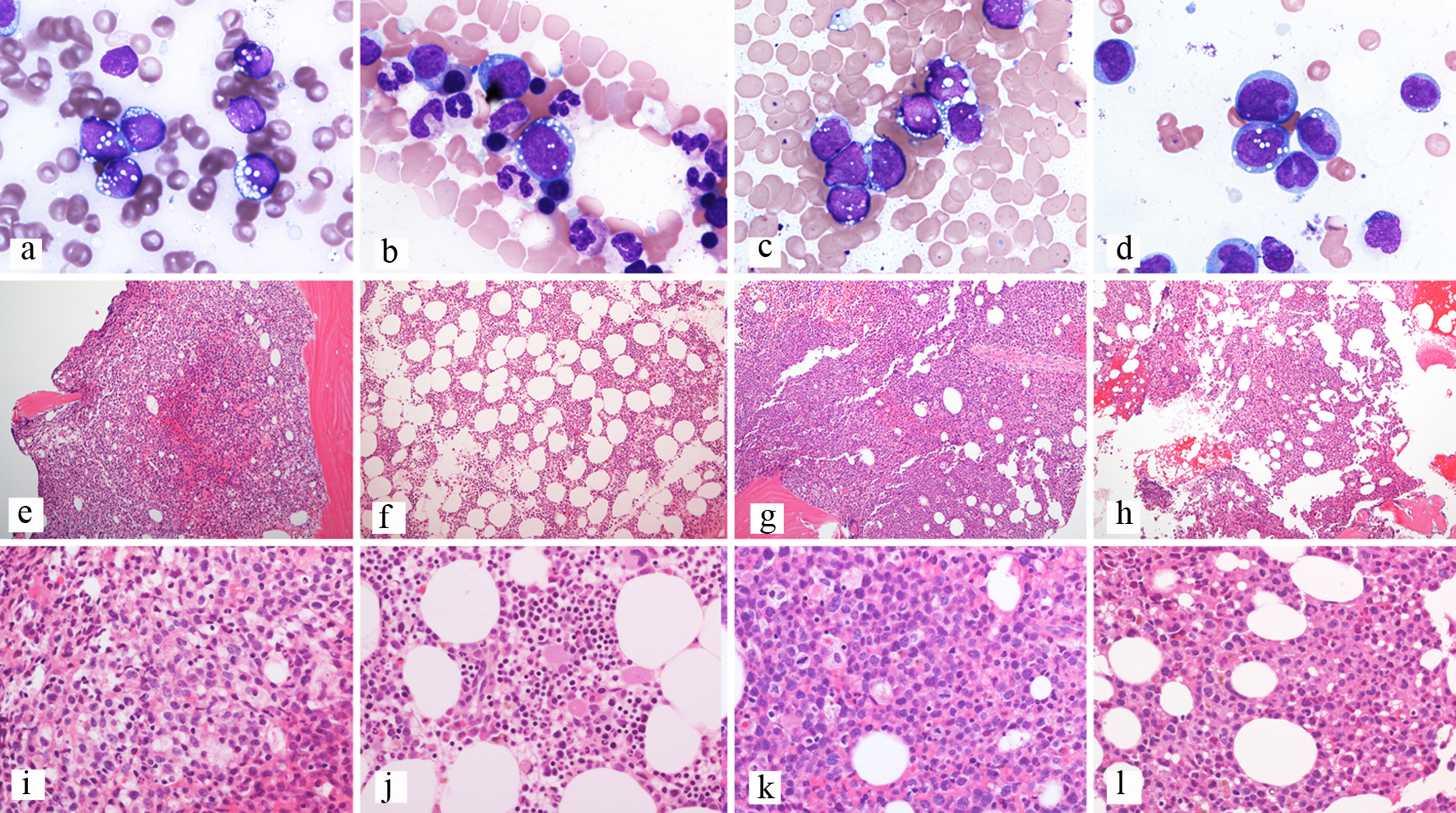
Figure 1. Composite of bone marrow biopsies from patient 3. (a, e, i) Bone marrow biopsy at diagnosis; (a) Aspirate showing blasts with cytoplasmic vacuoles, Wright-Giemsa stain, 100 × objective; (e) Trephine biopsy showing a hypercellular bone marrow, H&E-stained section, 10 × objective; (i) Trephine biopsy showing sheets of blasts, H&E-stained section, 40 × objective. (b, f, j) Bone marrow biopsy after enasidenib; (b) Aspirate showing rare erythroid precursors with cytoplasmic vacuoles and no definite blasts, Wright-Giemsa stain, 100 × objective; (f) Trephine biopsy showing a hypercellular bone marrow, H&E-stained section, 10 × objective; (j) Trephine biopsy showing trilineage hematopoiesis with erythroid hyperplasia, H&E-stained section, 40 × objective. (c, g, k) Bone marrow biopsy at relapse; (c) Aspirate showing blasts with cytoplasmic vacuoles, Wright-Giemsa stain, 100 × objective; (g) Trephine biopsy showing a hypercellular bone marrow, H&E-stained section, 10 × objective; (k) Trephine biopsy showing sheets of blasts, H&E-stained section, 40 × objective. (d, h, l) Bone marrow biopsy after enasidenib; (d) Aspirate showing blasts with partial differentiation and similar cytoplasmic vacuoles, Wright-Giemsa stain, 100 × objective; (h) Trephine biopsy showing a hypercellular bone marrow, H&E-stained section, 10 × objective; (l) Trephine biopsy showing sheets of immature cells, H&E-stained section, 40 × objective. H&E: hematoxylin and eosin.