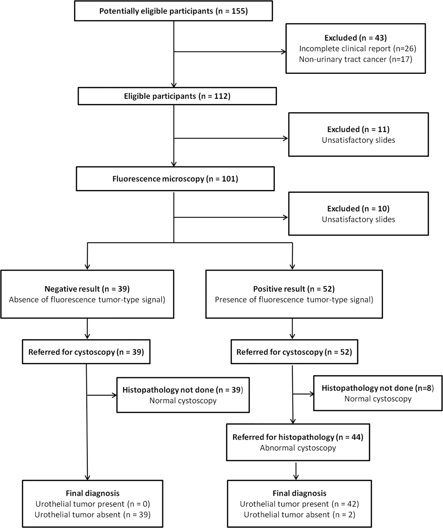
Figure 1. Diagram of STARD.
| World Journal of Oncology, ISSN 1920-4531 print, 1920-454X online, Open Access |
| Article copyright, the authors; Journal compilation copyright, World J Oncol and Elmer Press Inc |
| Journal website https://www.wjon.org |
Original Article
Volume 11, Number 5, October 2020, pages 204-215
Fluorescence Emitted by Papanicolaou-Stained Urothelial Cells Improves Sensitivity of Urinary Conventional Cytology for Detection of Urothelial Tumors
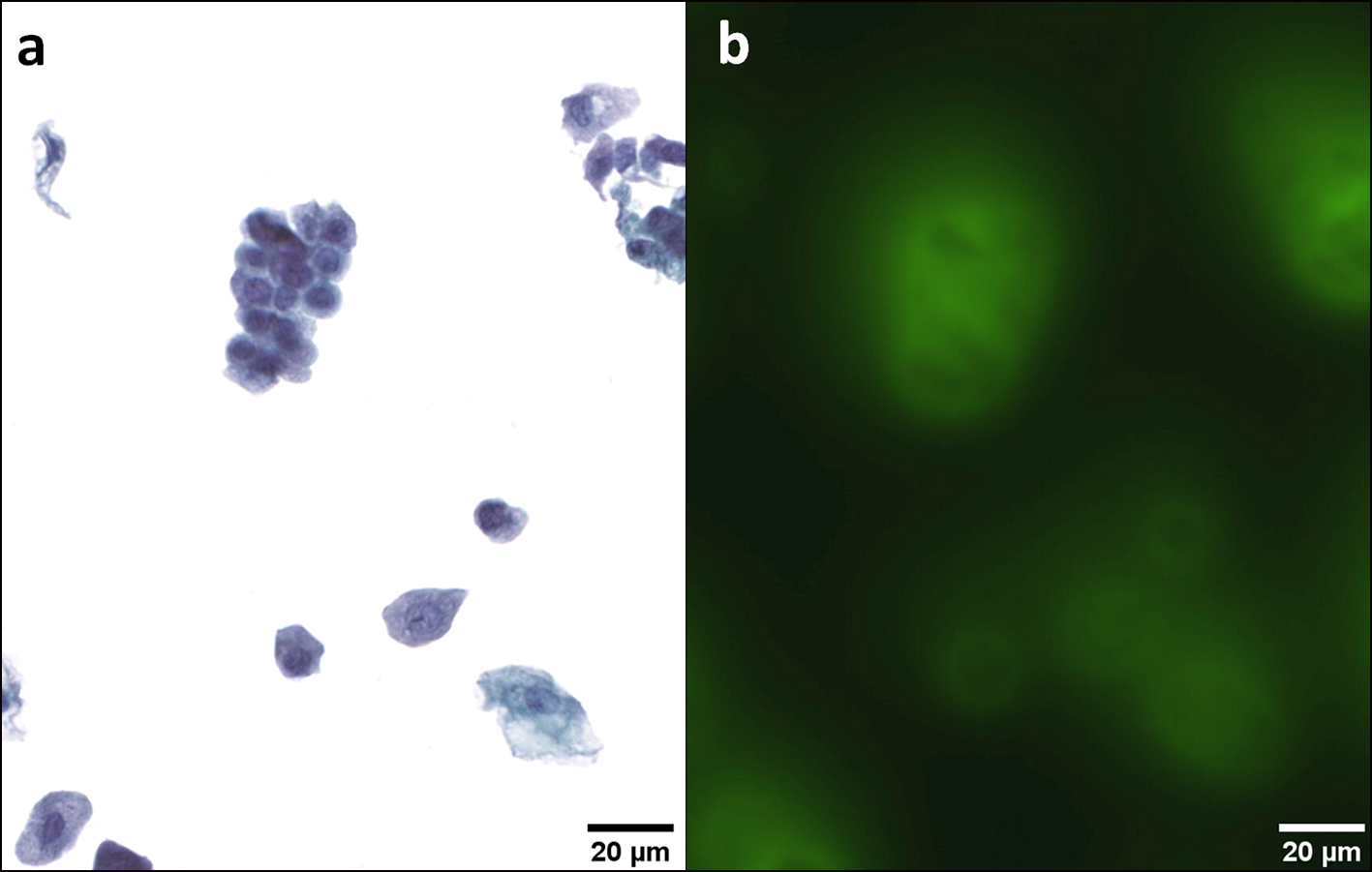
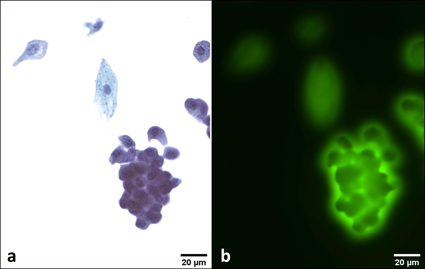
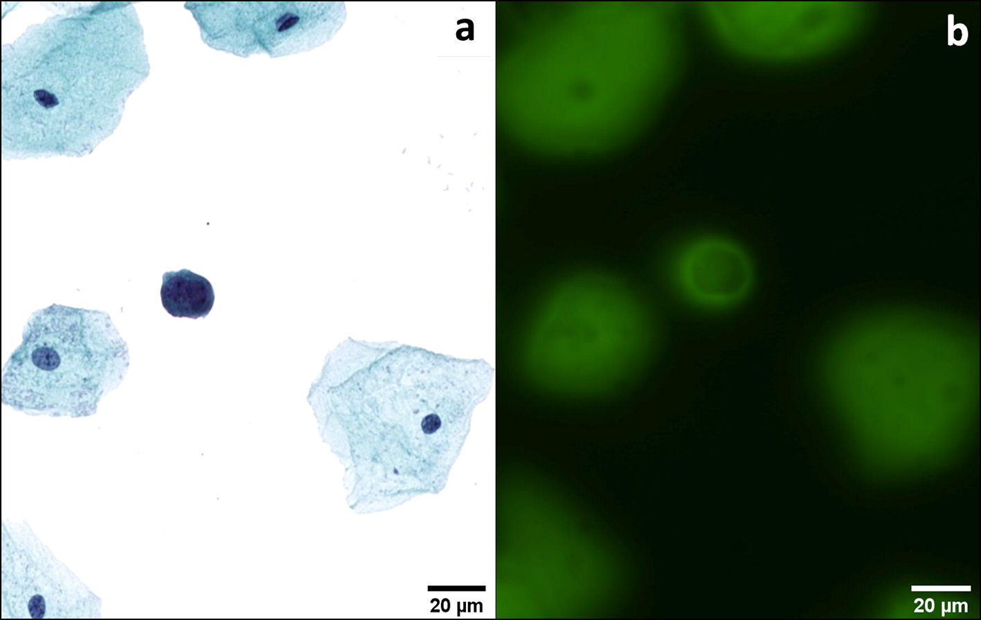
Figures

Tables
| Clinical diagnosis | FMi result +/- | UCCy | No. of cases | Follow-up time | |||||||
|---|---|---|---|---|---|---|---|---|---|---|---|
| TPS | Result +/- | ||||||||||
| HGUC (n = 16) | + | HGUC | + | 11 | NR | ||||||
| + | SHGUC | + | 1 | NR | |||||||
| + | AUC | + | 2 | NR | |||||||
| + | LGUN | + | 1 | NR | |||||||
| + | Negative | - | 1 | NR | |||||||
| LGUN (n = 26) | + | LGUN | + | 7 | NR | ||||||
| + | AUC | + | 5 | NR | |||||||
| + | Negative | - | 14 | NR | |||||||
| No tumor (n = 49) | - | Negative | - | 36 | No follow-up | ||||||
| - | AUC | + | 3 | No tumor (three cases), 6 - 60 months | |||||||
| + | Negative | - | 6 | No follow-up (one case), no tumor (five cases), 6 - 72 months | |||||||
| +a | SHGUC | +a | 1 | CIS 18 months | |||||||
| + | AUC | + | 3 | No tumor (three cases), 6 - 72 months | |||||||
| Statistics | FMi | UCCy | P* (FMi vs. UCCy) | FMi + UCCy | P* (FMi + UCCy vs. UCCy) | ||||||
| aCIS diagnosed after 18 months. *Significant threshold: P < 0.05. AUC: atypical urothelial cell; CIS: carcinoma in situ; FMi: fluorescence microscopy; HGUC: high-grade urothelial carcinoma; LGUN: low-grade urothelial neoplasia; NPV: negative predictive value; PPV: positive predictive value; SHGUC: suspicious for high-grade urothelial carcinoma; TPS: The Paris System; UCCy: urinary conventional cytology. | |||||||||||
| HGUC + LGUN | |||||||||||
| Sensitivity, % | 100 | 64.3 | 0.0001* | 100 | 0.0001* | ||||||
| Specificity, % | 79.6 | 85.7 | 0.3173 | 73.47 | 0.0143* | ||||||
| PPV, % | 80.8 | 79.4 | 0.8238 | 76.36 | 0.5051 | ||||||
| NPV, % | 100 | 73.7 | 0.0001* | 100 | 0.0001* | ||||||
| HGUC | |||||||||||
| Sensitivity, % | 100 | 93.7 | 0.3173 | ||||||||
| LGUC | |||||||||||
| Sensitivity, % | 100 | 46.2 | 0.0002* | ||||||||
| Cystoscopy | Clinical diagnosis | No. of cases | FMi result +/- | UCCy | Follow-up time | ||||||
|---|---|---|---|---|---|---|---|---|---|---|---|
| 2016 WHO | Stage | TPS | Result +/- | ||||||||
| Positive | LGUC | pTa | 6 | + | Negative | - | IR | ||||
| Positive | LGUC | pTa | 2 | + | AUC | + | IR | ||||
| Positive | LGUC | pTa | 4 | + | LGUN | + | IR | ||||
| Positive | LGUC | pT1 | 1 | + | AUC | + | IR | ||||
| Positive | LGUC | pT1 | 1 | + | LGUN | + | IR | ||||
| Positive | LGUC | pT1 | 3 | + | Negative | - | IR | ||||
| Positive | HGUC | pTa | 1 | + | LGUN | + | IR | ||||
| Positive | HGUC | pTa | 1 | + | HGUC | + | IR | ||||
| Positive | HGUC | pT1 | 1 | + | AUC | + | IR | ||||
| Positive | HGUC | pT1 | 1 | + | HGUC | + | IR | ||||
| Positive | HGUC | pT2 | 1 | + | Negative | - | IR | ||||
| n = 22 | |||||||||||
| Negative | No disease | 8 | - | Negative | - | No follow-up | |||||
| Inflammationa | No tumor | 1 | + | AUC | + | No tumor, 36 months | |||||
| Negativeb | No tumor | 1 | + | SHGUC | + | CIS, 18 months | |||||
| Negative | No tumor | 1 | + | AUC | + | No tumor, 6 months | |||||
| n = 11 | |||||||||||
| Statistics | FMi | UCCy | P* | ||||||||
| aBCG therapy. bBCG therapy and CIS after 18 months. *Significant threshold: P < 0.05. AUC: atypical urothelial cell; CIS: carcinoma in situ; FMi: fluorescence microscopy; HGUC: high-grade urothelial carcinoma; IR: irrelevant; LGUC: low-grade urothelial carcinoma; LGUN: low-grade urothelial neoplasia; NPV: negative predictive value; PPV: positive predictive value; SHGUC: suspicious for high-grade urothelial carcinoma; TPS: The Paris System; UCCy: urinary conventional cytology; WHO: World Health Organization. | |||||||||||
| Sensitivity, % | 100 | 54.5 | 0.0016* | ||||||||
| Specificity, % | 72.7 | 72.7 | NA | ||||||||
| PPV, % | 88 | 80 | 0.0021* | ||||||||
| NPV, % | 100 | 44.4 | 0.1289 | ||||||||
| Cystoscopy | Clinical diagnosis | No. of cases | FMi result +/- | UCCy | Follow-up time | ||||||
|---|---|---|---|---|---|---|---|---|---|---|---|
| 2016 WHO | Stage | TPS | Result +/- | ||||||||
| Positive | LGUC | pTa | 4 | + | Negative | - | IR | ||||
| Positive | LGUC | pTa | 2 | + | AUC | + | IR | ||||
| Positive | LGUC | pTa | 2 | + | LGUN | + | IR | ||||
| Negativea | LGUC | pTx | 1 | + | Negative | - | IR | ||||
| Positive | HGUC | pTa | 3 | + | HGUC | + | IR | ||||
| Positive | HGUC | pT1 | 1 | + | SHGUC | + | IR | ||||
| Positive | HGUC | pT1 | 1 | + | HGUC | + | IR | ||||
| Positive | HGUC | pT2 | 1 | + | AUC | + | IR | ||||
| Positive | HGUC | pT2 | 1 | + | HGUC | + | IR | ||||
| Positive | HGUC | pT3 | 2 | + | HGUC | + | IR | ||||
| Positive | HGUC | pT4 | 2 | + | HGUC | + | IR | ||||
| n = 20 | |||||||||||
| Negative | No disease | 14 | - | Negative | - | No follow-up | |||||
| Negative | BPH | 5 | - | Negative | - | No follow-up | |||||
| Negative | Lithiasis | 3 | - | Negative | - | No follow-up | |||||
| Negative | Infection | 4 | - | Negative | - | No follow-up | |||||
| Negative | Inflammation | 2 | - | Negative | - | No follow-up | |||||
| Abnormalb | BPH + inflammation | 1 | + | Negative | - | No tumor, 6 months | |||||
| Negative | BPH + inflammation | 1 | + | Negative | - | No tumor, 72 months | |||||
| Negative | Prostatitis | 2 | + | Negative | - | No tumor, 12 and 24 months | |||||
| Negative | Lithiasis | 2 | + | Negative | - | No tumor, 0 and 12 months | |||||
| Negative | Lithiasis | 1 | - | AUC | + | No tumor, 60 months | |||||
| Negative | Inflammation | 2 | - | AUC | + | No tumor, 6 and 24 months | |||||
| Abnormalb | Infection | 1 | + | AUC | + | No tumor, 72 months | |||||
| n = 38 | |||||||||||
| Statistics | FMi | UCCy | P* | ||||||||
| aUreteral urothelial tumor. bDiagnosis confirmed by bladder biopsy. *Significant threshold: P < 0.05. AUC: atypical urothelial cell; BPH: benign prostatic hyperplasia; FMi: fluorescence microscopy; HGUC: high-grade urothelial carcinoma; IR: irrelevant; LGUC: low-grade urothelial carcinoma; LGUN: low-grade urothelial neoplasia; NPV: negative predictive value; PPV: positive predictive value; SHGUC: suspicious for high-grade urothelial carcinoma; UCCy: urinary conventional cytology; WHO: World Health Organization. | |||||||||||
| Sensitivity, % | 100 | 75 | 0.0253* | ||||||||
| Specificity, % | 81.6 | 89.5 | 0.3173 | ||||||||
| PPV, % | 74.1 | 78.9 | 0.6314 | ||||||||
| NPV, % | 100 | 87.2 | 0.0255* | ||||||||