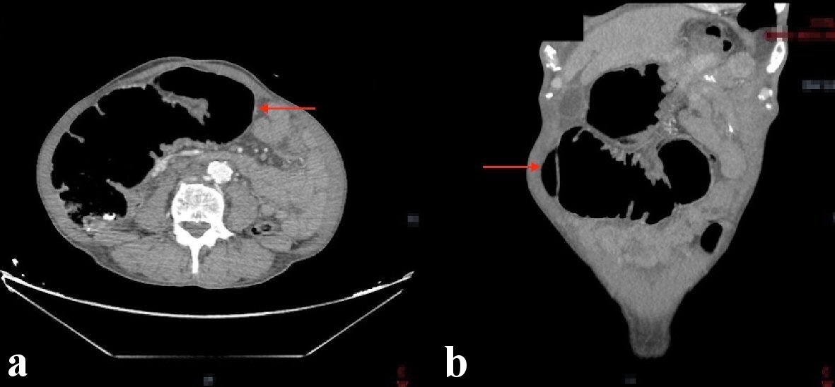
Figure 1. CT abdomen showing a mid-transverse colon mass measuring 30 × 44 mm resulting in proximal colonic obstruction (red arrows). (a) Axial view. (b) Coronal view. CT: computed tomography.
| World Journal of Oncology, ISSN 1920-4531 print, 1920-454X online, Open Access |
| Article copyright, the authors; Journal compilation copyright, World J Oncol and Elmer Press Inc |
| Journal website https://www.wjon.org |
Case Report
Volume 12, Number 4, August 2021, pages 127-131
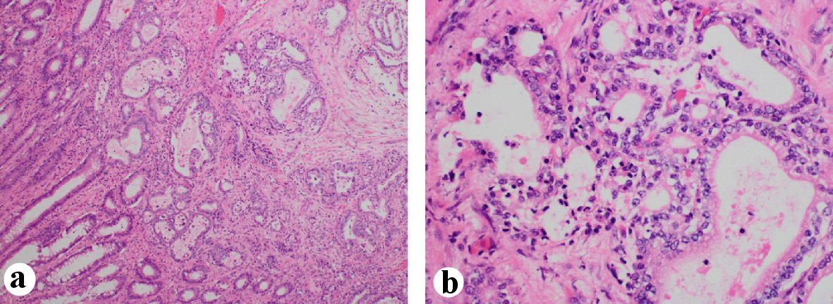
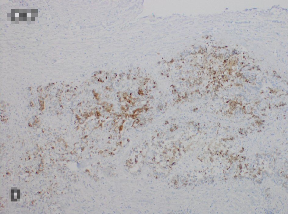
Metastatic Gastric Cancer to the Colon
Figures