Figures
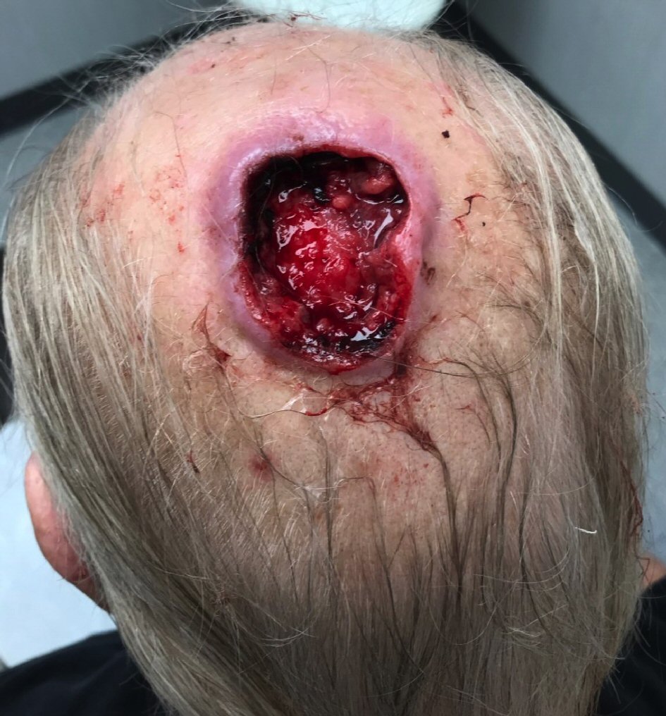
Figure 1. A representative image of the scalp mass of the presented patient. A large, ulcerated mass on the left scalp was noted, which was mobile and not fixated to the scalp.
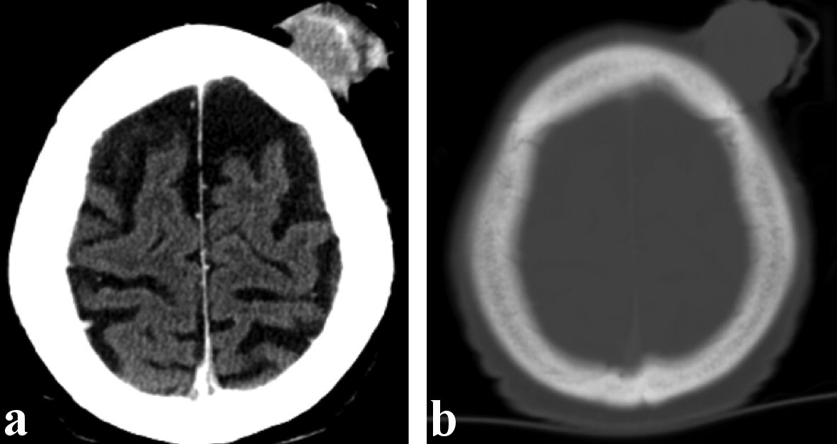
Figure 2. Representative computed tomography (CT) images of the presented patient. (a) Soft tissue window demonstrating a mildly enhancing exophytic left frontal scalp mass measuring 3.4 × 3.3 cm. (b) Bone window demonstrating no evidence of invasion to the underlying bone.
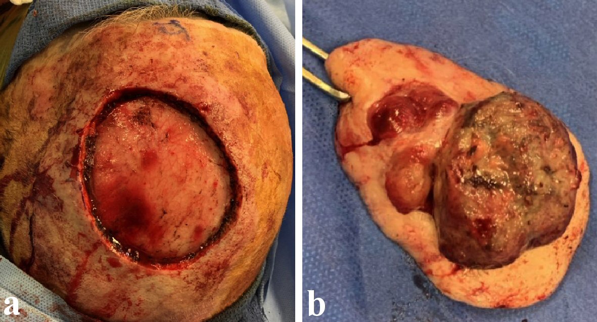
Figure 3. Representative images of the intraoperative findings of the presented patient. (a) Wide local excision of the scalp lesion was performed and the pericranium of the scalp was resected as a deep margin. (b) The gross finding of the specimen, which measured 5.4 × 5.1 × 2.2 cm.
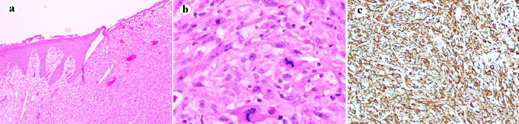
Figure 4. Pathology slides of the scalp leiomyosarcoma in the presented patient. (a) Low-power field (× 10): skin with ulceration and proliferation of neoplastic smooth muscles cells occupying the dermis. (b) High-power field (× 40): neoplastic smooth muscle cells showing cytologic atypia, frequent mitosis and single cell apoptosis. (c) Medium-power field (× 20) with immunostain for desmin: the tumor is diffusely positive with desmin.