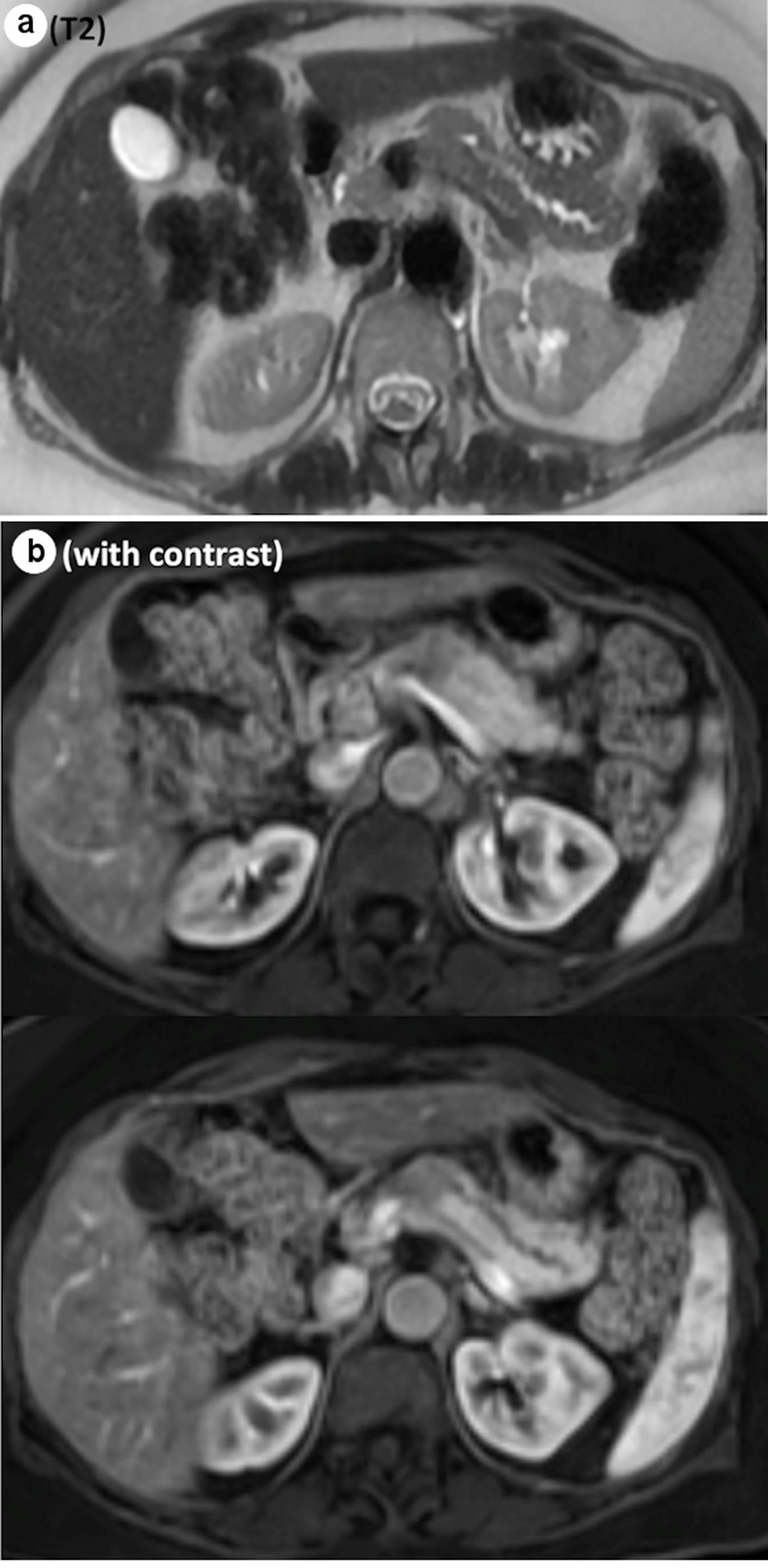
Figure 1. Representative images from MRI. (a) Abrupt cut off of the main pancreatic duct on the T2 sequence. (b) Hypoenhancing mass, measuring 2.4 × 1.9 × 3.2 cm, on the contrast study. MRI: magnetic resonance imaging.
| World Journal of Oncology, ISSN 1920-4531 print, 1920-454X online, Open Access |
| Article copyright, the authors; Journal compilation copyright, World J Oncol and Elmer Press Inc |
| Journal website https://www.wjon.org |
Case Report
Volume 12, Number 6, December 2021, pages 240-245
Immunoglobulin G4-Negative Inflammatory Pseudotumors of the Pancreas
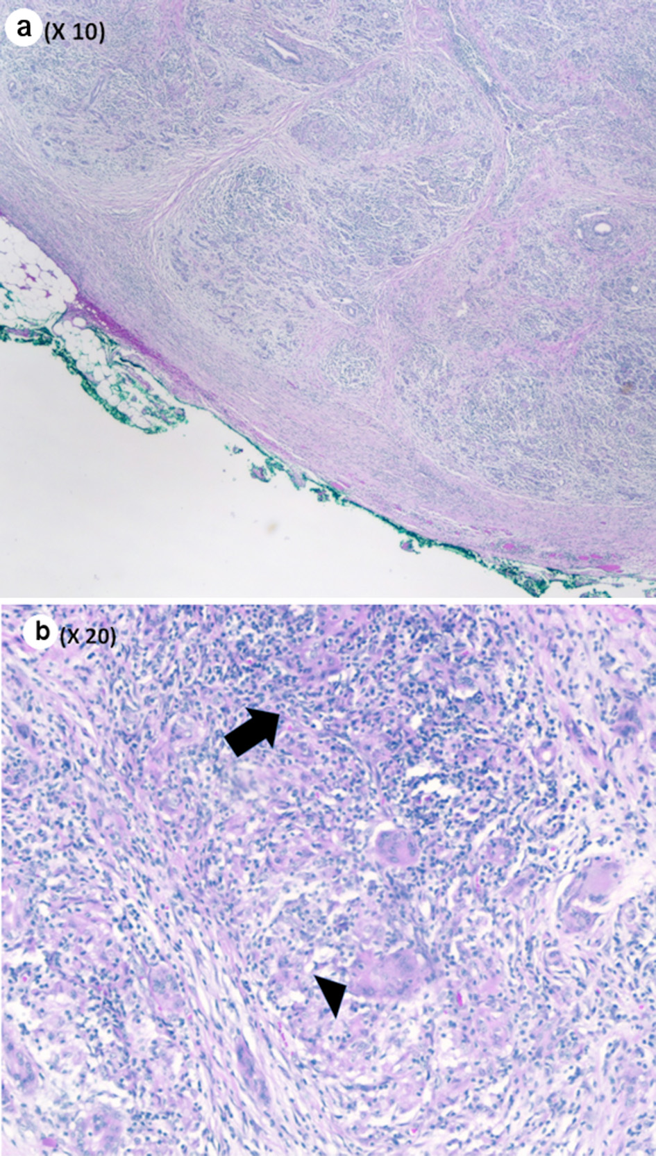
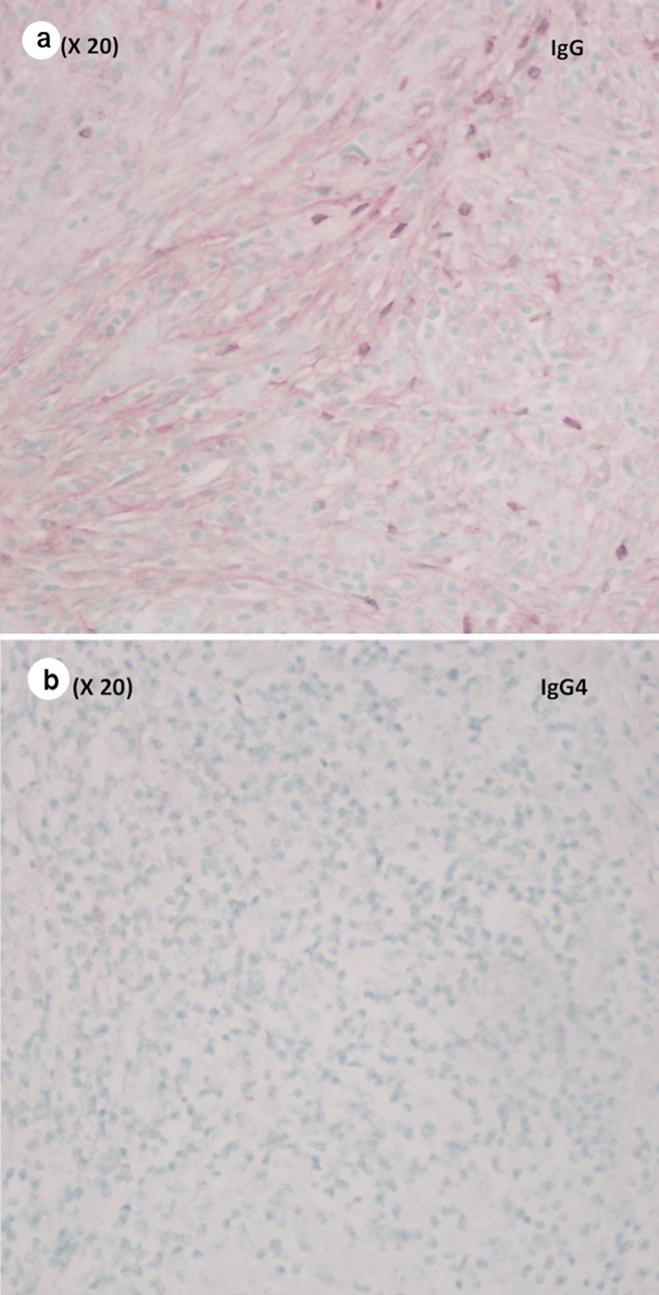
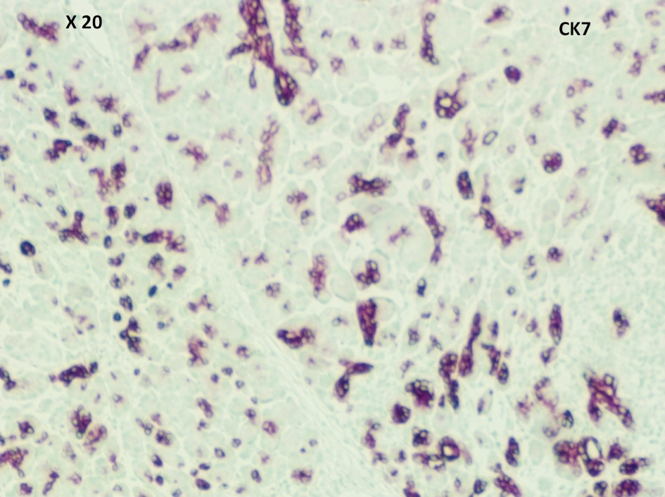
Figures