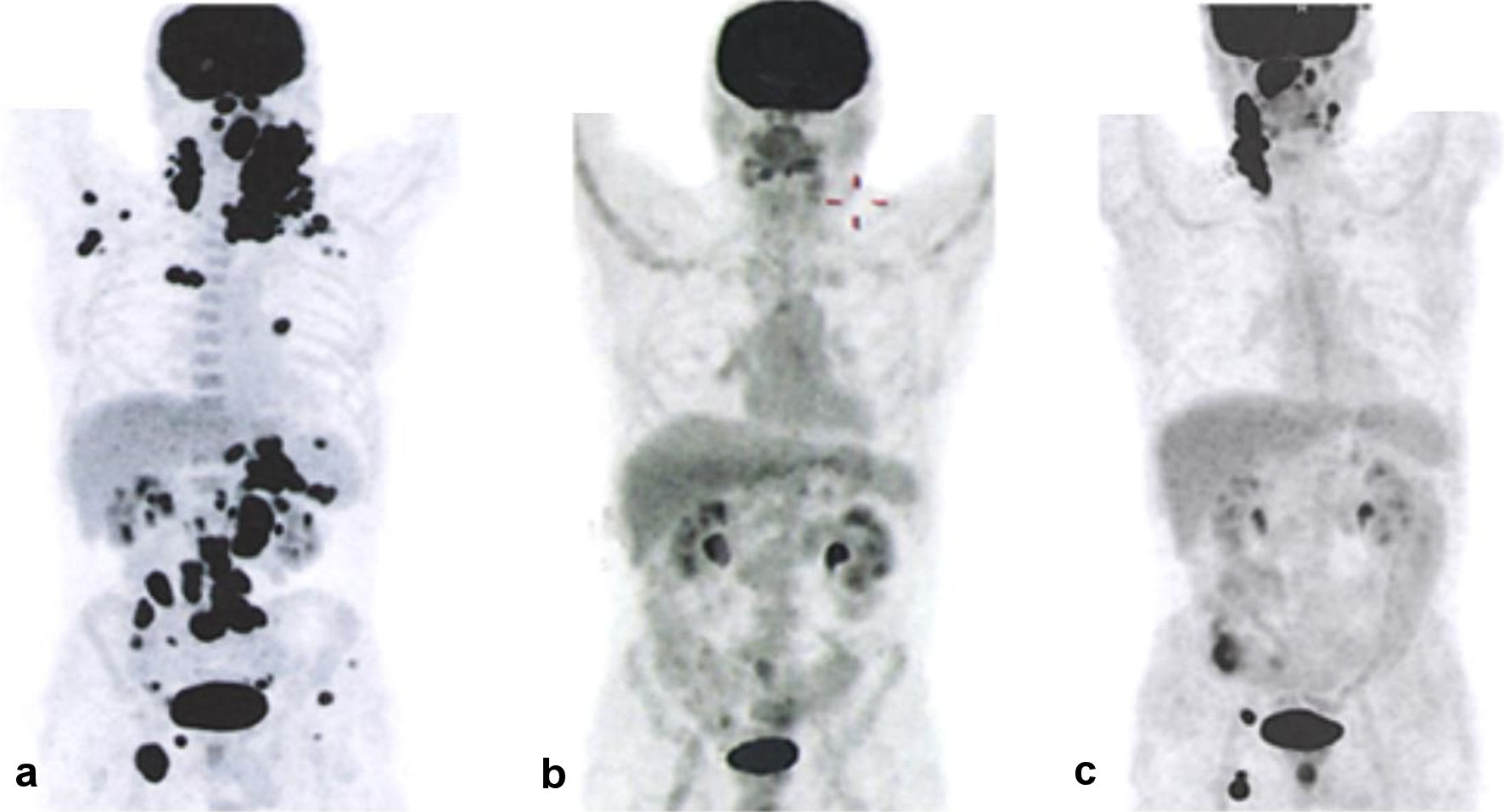
Figure 1. PET/CT results showing disease progression. (a) Before the start of therapy. (b) After the end of therapy. (c) Following disease relapse. PET/CT: positron emission tomography combined with computed tomography.
| World Journal of Oncology, ISSN 1920-4531 print, 1920-454X online, Open Access |
| Article copyright, the authors; Journal compilation copyright, World J Oncol and Elmer Press Inc |
| Journal website https://www.wjon.org |
Case Report
Volume 13, Number 1, February 2022, pages 38-47
Non-GCB Diffuse Large B-Cell Lymphoma With an Atypical Disease Course: A Case Report and Clinical Exome Analysis
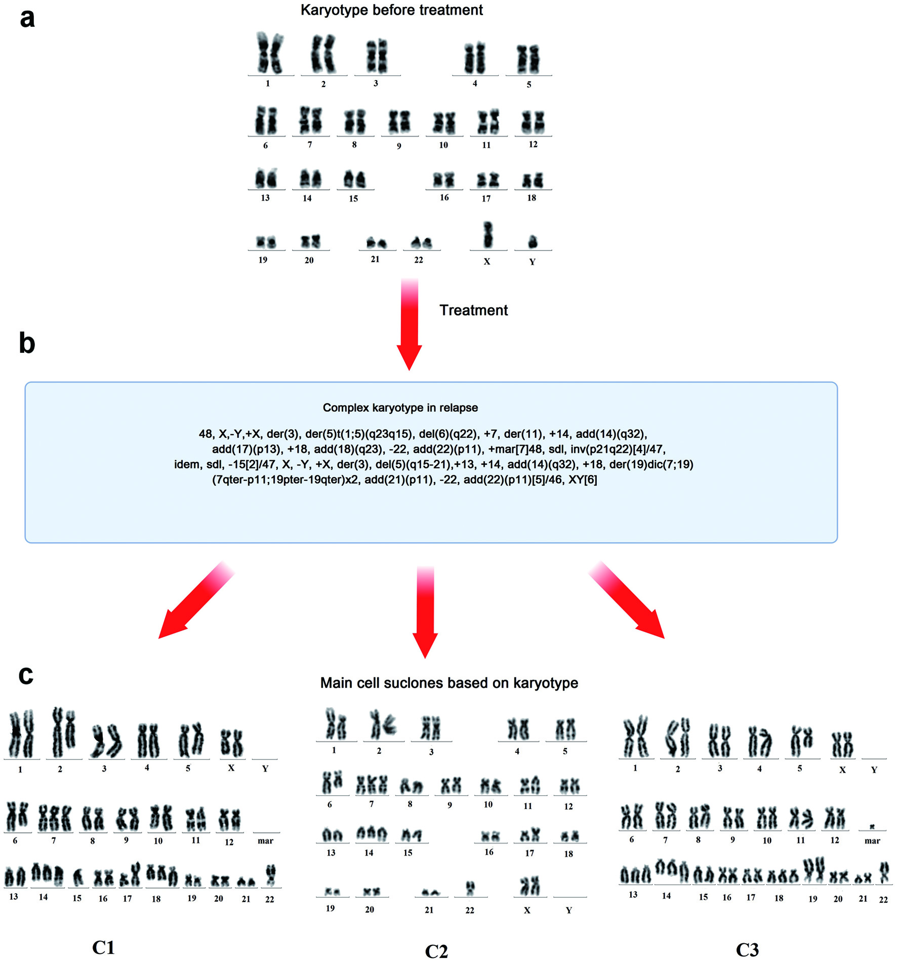
Figures