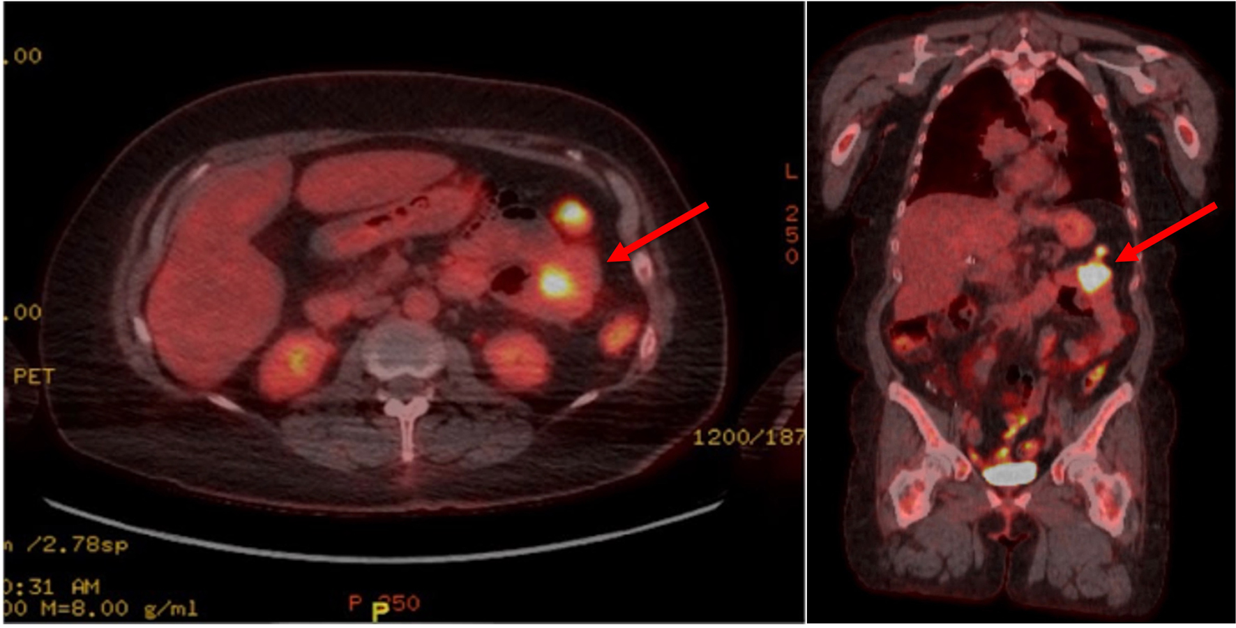
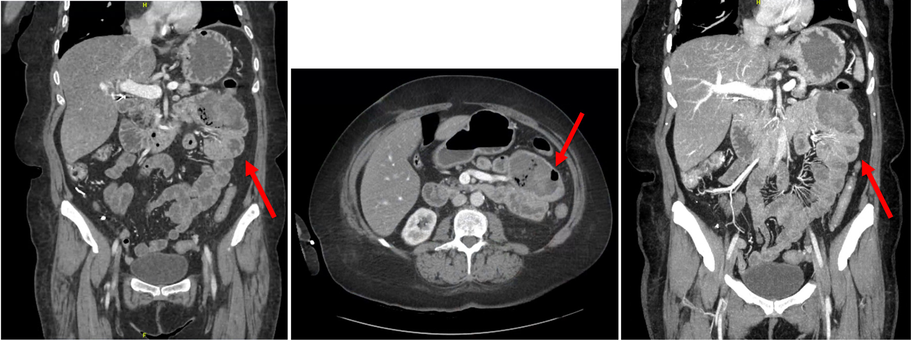
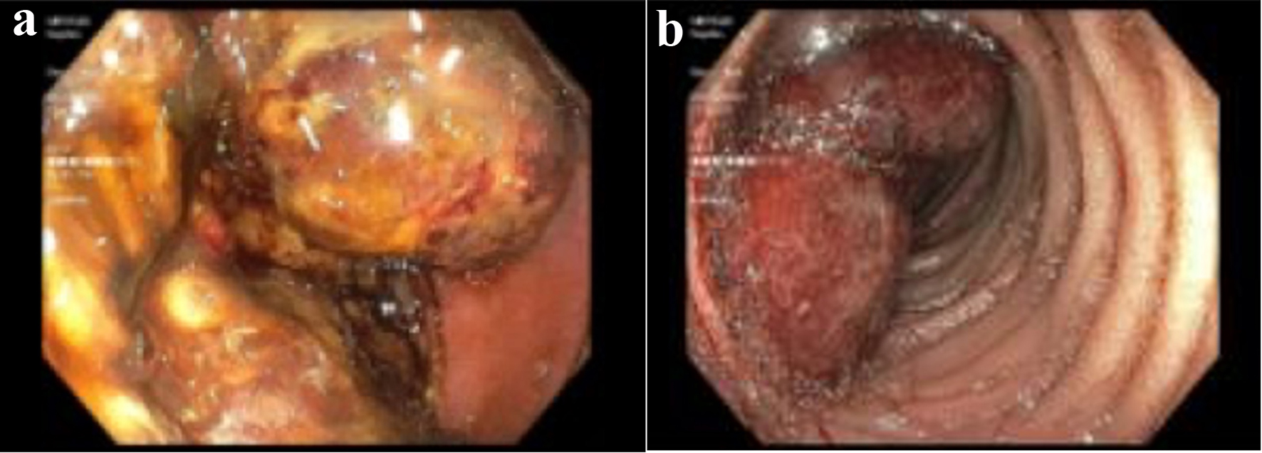
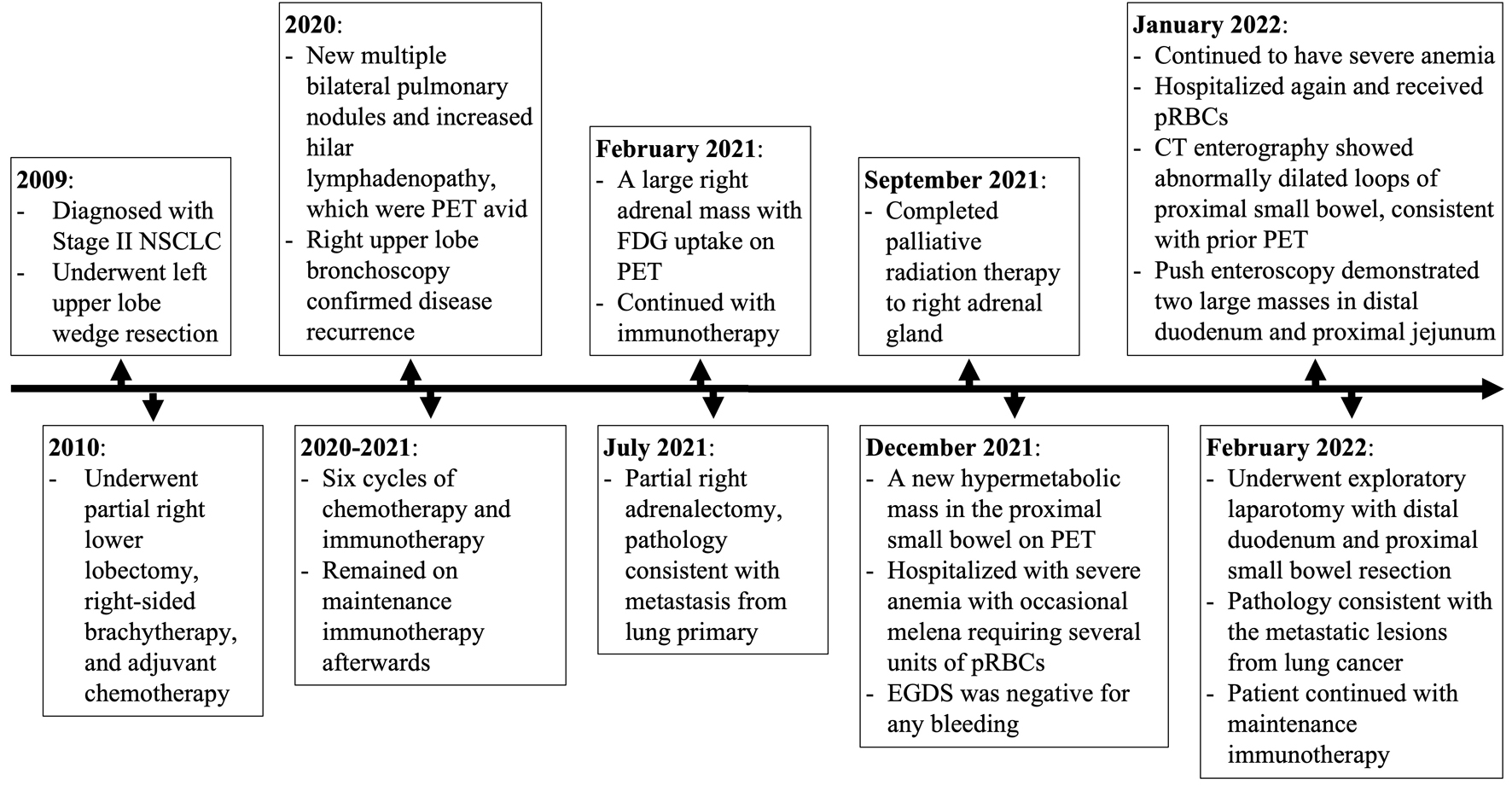
| Ogasawara et al (2022) [24] | 62 M | Adenocarcinoma | Abdominal pain, anemia, about 7 cm tumor in left abdomen on CT | Jejunum | Biopsy from enteroscopy |
| Suzuki et al (2022) [25] | 70 M | Pleomorphic carcinoma | Generalized fatigue, abdominal pain, anemia, CT showed large mass in left abdominal cavity, endoscopy showed tumor in jejunum | Jejunum | Biopsy from endoscopy and surgical resection |
| Kang et al (2021) [15] | 66 M | Adenocarcinoma | Melena, dizziness, anemia, 2 cm polypoidal lesion in second portion of duodenum | Total of 22 lesions in stomach, duodenum, jejunum, and ileum - ranging from 0.8 to 4.0 cm | Biopsy from EGDS and surgical resections |
| O’Neill et al (2020) [26] | 56 M | Adenocarcinoma | Headache and dysarthria from lung mass, small bowel mass noted on PET with no attributable symptoms | Second portion of duodenum | Biopsy from gastroscopy |
| Xie et al (2020) [27] | 55 M | Pulmonary sarcomatoid carcinoma | Epigastric pain, melena, anemia, PET showed wall thickening of small bowel | Duodenum and jejunum | Surgical resection |
| Xie et al (2020) [27] | 61 M | Pulmonary sarcomatoid carcinoma | Melena, intermittent fevers, anemia, PET showed circumferential thickening of small intestines | Small bowel - not fully described | Biopsy from gastroscopy |
| Plestina et al (2019) [28] | 57 M | Undifferentiated pleomorphic sarcoma | Acute abdomen secondary to bowel perforation, CT scan 3 months after lung surgery showed a 60 mm obstructive metastatic small bowel lesion | Ileum | Surgical resection |
| Chen et al (2018) [29] | 69 M | Adenocarcinoma | Bloating, decreased flatus, intestinal effusion and obstruction on CT | Small bowel - not fully described | Surgical resection |
| Ohira et al (2018) [30] | 75 M | Adenocarcinoma | Melena, anemia, capsule endoscopy showed polyp in small bowel | Jejunal mass | Biopsy from enteroscopy and autopsy |
| Janez et al (2017) [31] | 53 M | Adenocarcinoma | Upper abdominal pain, vomiting, constipation, SBO on CT, CEA elevated | Multiple metastatic lesions along small bowel and small bowel mesentery | Surgical resection |
| Ying et al (2017) [32] | 59 M | Adenocarcinoma | Melena, anemia, fecal occult blood, CT and PET demonstrated small bowel mass | Jejunum | Surgical resection |


