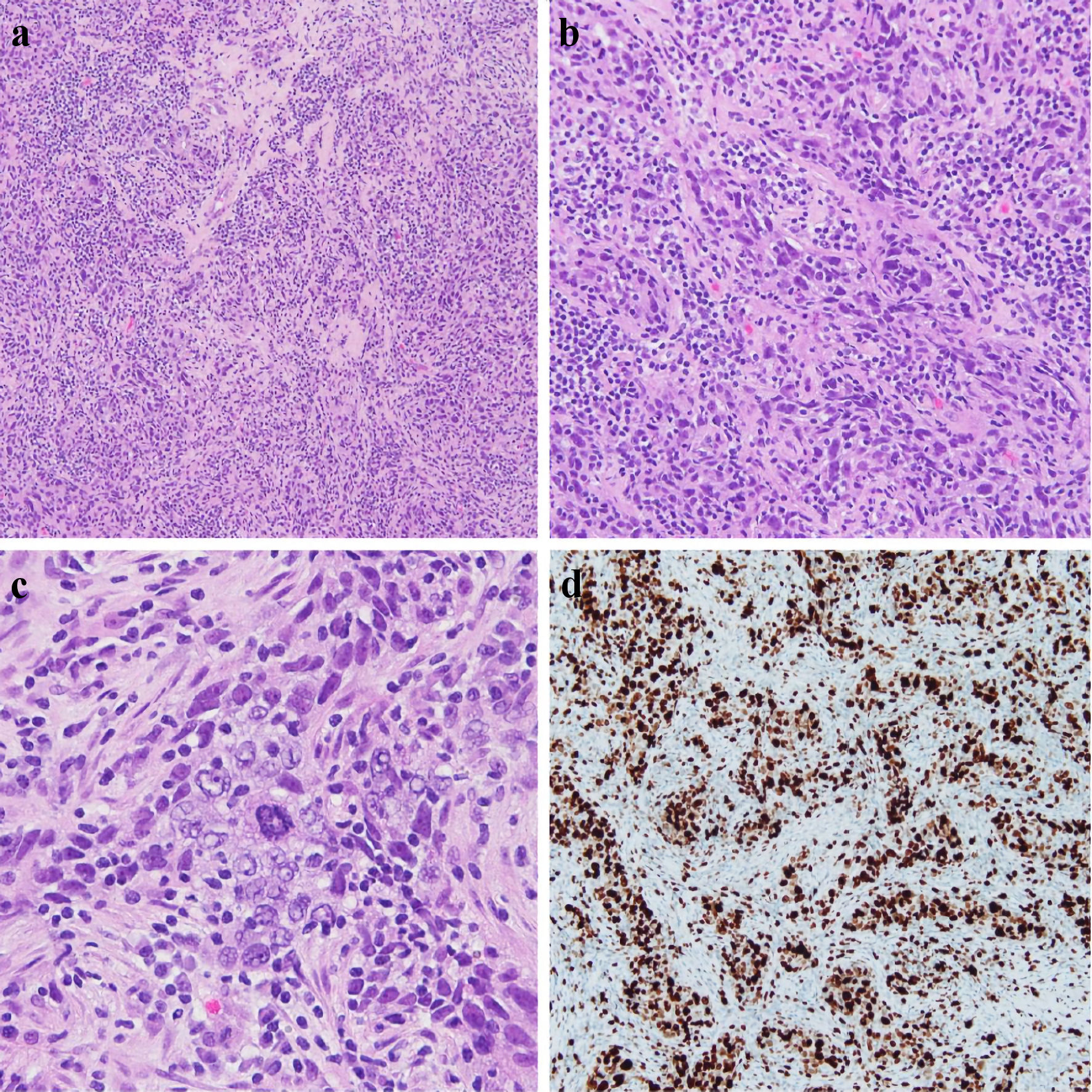
Figure 1. Right core breast biopsy (a, × 10), (b, × 20), (c, × 40) showing infiltrating tumor cells with large and atypical nuclei, a syncytial growth pattern, and a background of lymphocytes. An atypical mitotic figure is present in the center of (c). Ki67 IHC stain showing > 90% nuclear staining of tumor cells, reflecting increased proliferation (d). IHC: immunohistochemistry.
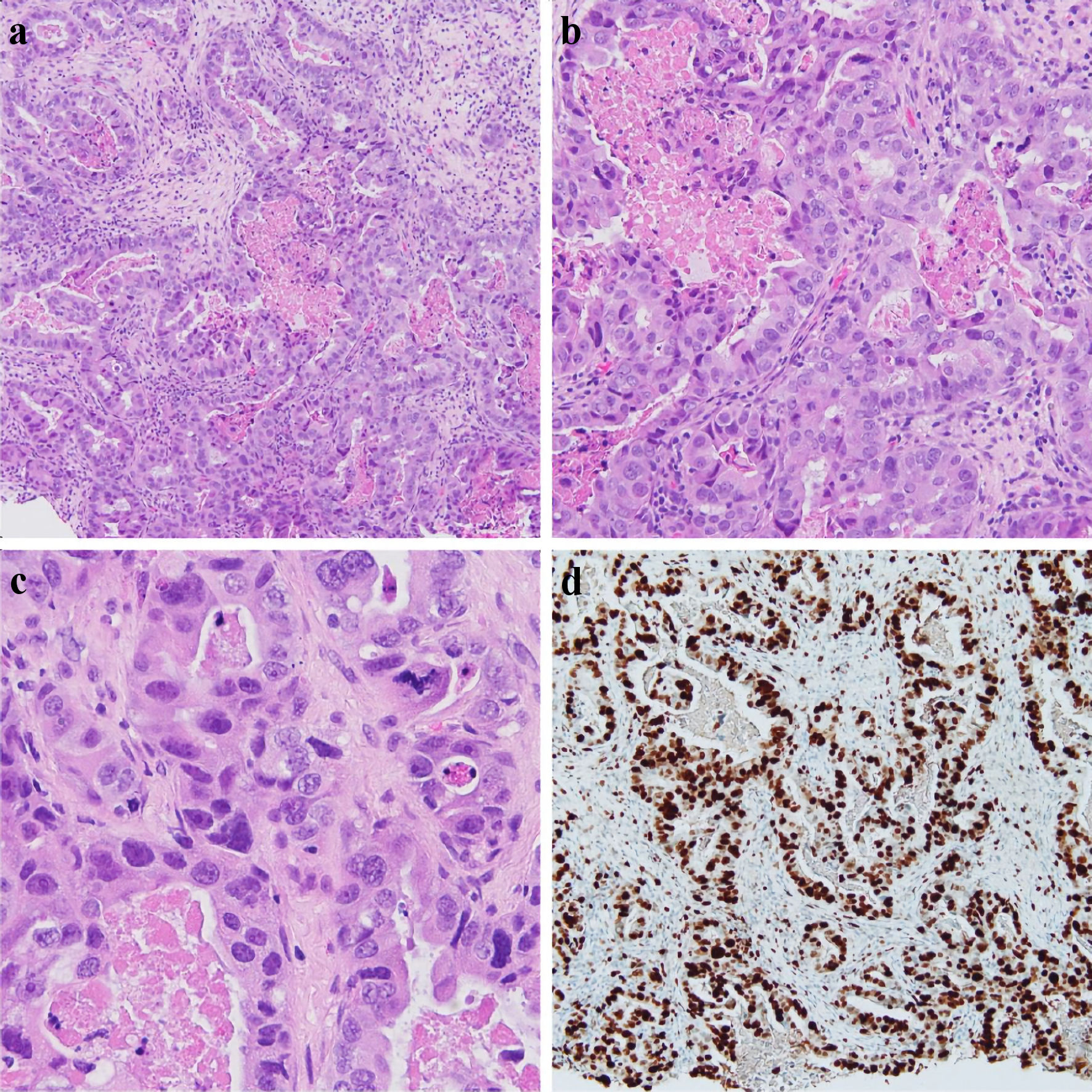
Figure 2. Left breast core biopsy (a, × 10), (b, × 20), (c, × 40) showing infiltrating tumor cells with large, pleomorphic nuclei, with a glandular/tubule formation and associated necrosis within the tubules. An atypical mitotic figure can be seen in (c). Ki67 IHC stain showing > 90% nuclear staining of tumor cells, reflecting increased proliferation (d). IHC: immunohistochemistry.