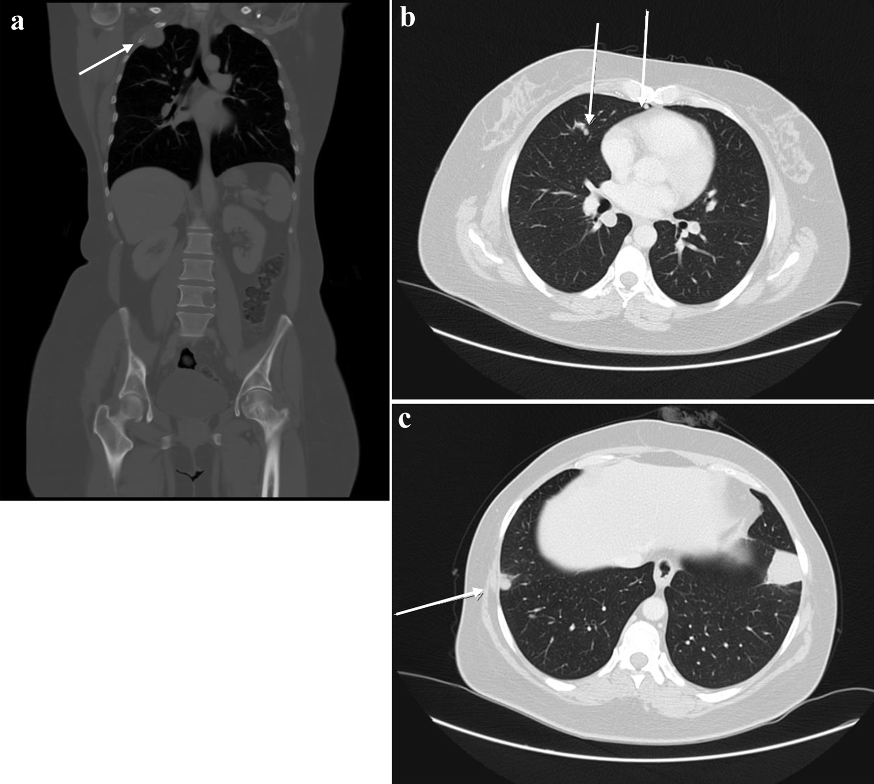
Figure 2. First disease progression shown on CT scan with contrast, manifested with multiple bilateral pulmonary nodules and two prominent right hilar lymph nodes, with multiple scattered destructive lytic bony lesions (arrows) (August 12, 2020) (a: coronal, bone window; b, c: axial, lung window). CT: computed tomography.
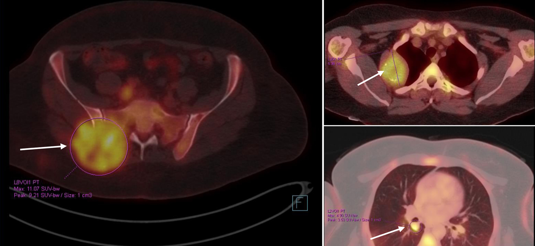
Figure 3. PET/CT scan showing widespread metastatic disease (arrows) (September 10, 2020). Beta-hCG level was within normal levels. Beta-hCG: beta human chorionic gonadotropin; CT: computed tomography; PET: positron emission tomography.
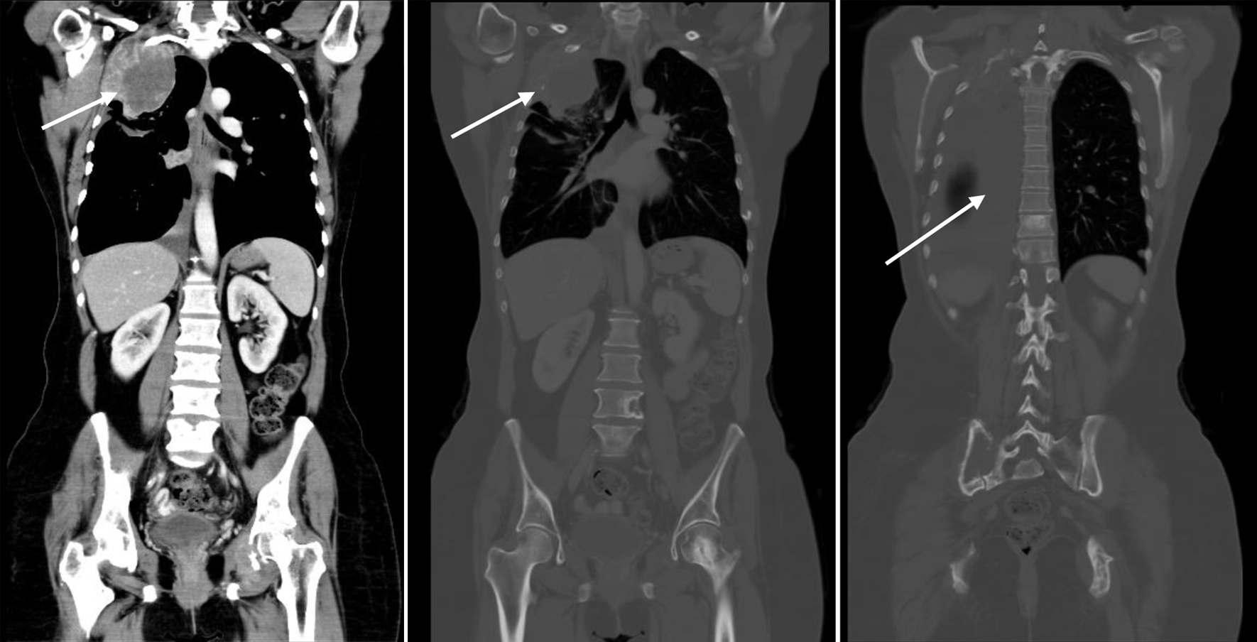
Figure 4. CT scan showing disease progression (February 9, 2023): enlarging extraosseous soft tissue component associated with destructive bony lesion involving the right second rib and left acetabulum, enlarging bilateral pulmonary lesions, and moderate right-sided pleural effusion with new right pleural nodular thickening, suspicious for pleural carcinomatosis (arrows). Beta-hCG level was elevated (8.43 mIU/mL). Beta-hCG: beta human chorionic gonadotropin; CT: computed tomography.