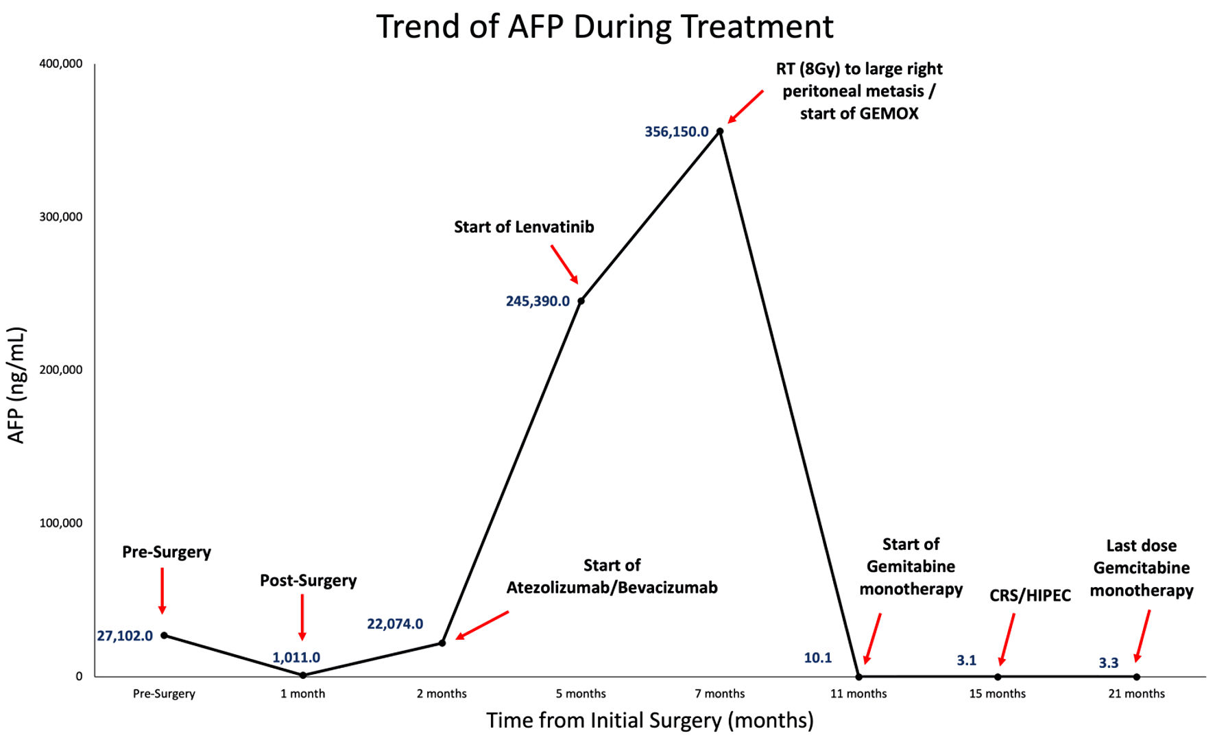
Figure 1. AFP trend starting from initial surgery to subsequent treatments (including atezolizumab/bevacizumab, lenvatinib, palliative radiation (RT) of 8 Gy to large right peritoneal metastasis, GEMOX, and gemcitabine monotherapy), CRS/HIPEC, and completion of postoperative gemcitabine therapy. GEMOX: gemcitabine and oxaliplatin; AFP: alpha fetoprotein; CRS: cytoreductive surgery; HIPEC: hyperthermic intraperitoneal chemotherapy.

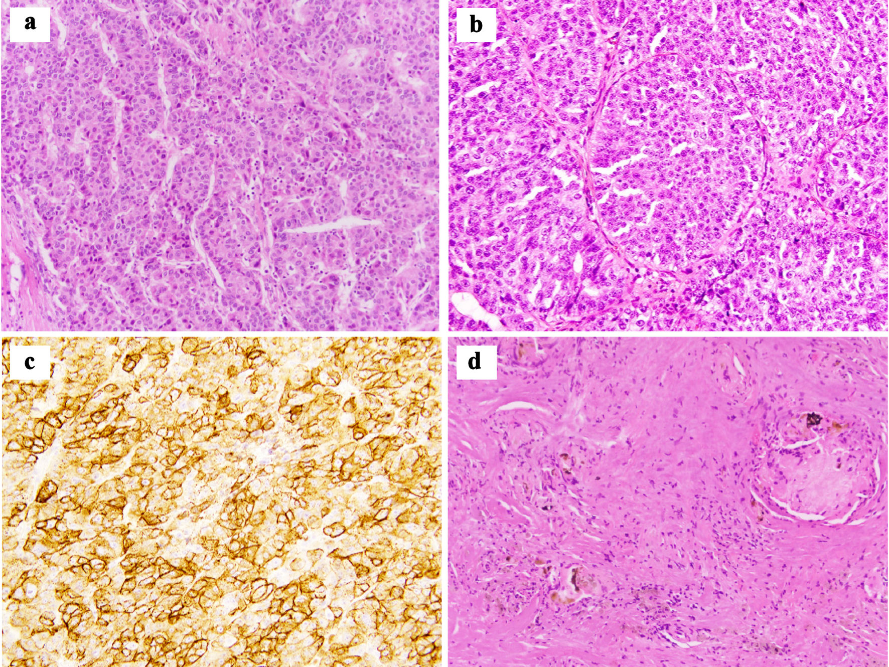
Figure 2. (a) Pathological analysis of initial tumor resection reveals moderately to poorly differentiated hepatocellular carcinoma with characteristic macrotrabecular growth pattern (hematoxylin and eosin, × 200). (b, c) Peritoneal nodule shows metastatic poorly differentiated hepatocellular carcinoma that stains diffusely positive for glypican 3 ((b) hematoxylin and eosin, × 200; (c) glypican 3, × 200). (d) Post-GEMOX resection specimen shows foci of necrosis and bile plugs surrounded by foreign body giant cell reaction with no viable tumor present (hematoxylin and eosin, × 200). GEMOX: gemcitabine and oxaliplatin.

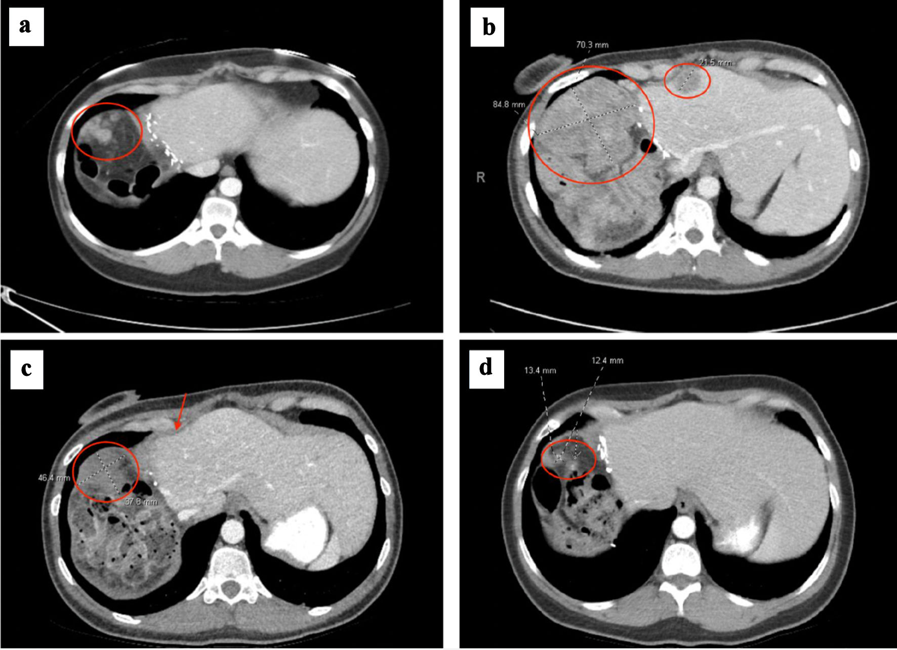
Figure 3. (a) Right upper quadrant peritoneal metastasis (circled) at time of metastatic recurrence after initial hepatectomy. (b) Right upper quadrant peritoneal metastasis (circled) increased to 8.5 cm, which had progressed after no response to atezolizumab/bevacizumab and lenvatinib. Smaller mid-anterior surface perihepatic implant also noted (circled). (c) Right upper quadrant peritoneal metastasis decreased to 4.6 cm (circled) and anterior perihepatic implant (arrow) resolved after 2 months of GEMOX. (d) Right upper quadrant peritoneal metastasis decreased to 1.3 cm prior to cytoreductive surgery and HIPEC, after several months of GEMOX followed by gemcitabine monotherapy. GEMOX: gemcitabine and oxaliplatin; HIPEC: hyperthermic intraperitoneal chemotherapy.


