World Journal of Oncology, ISSN 1920-4531 print, 1920-454X online, Open Access
Article copyright, the authors; Journal compilation copyright, World J Oncol and Elmer Press Inc
Journal website https://www.wjon.org

Original Article

Volume 000, Number 000, July 2024, pages 000-000


On the Origin of Abdominal Venous Leiomyosarcomas: The Role of the Sex-Hormone Drainage Pathways

Figures

Figure 1.
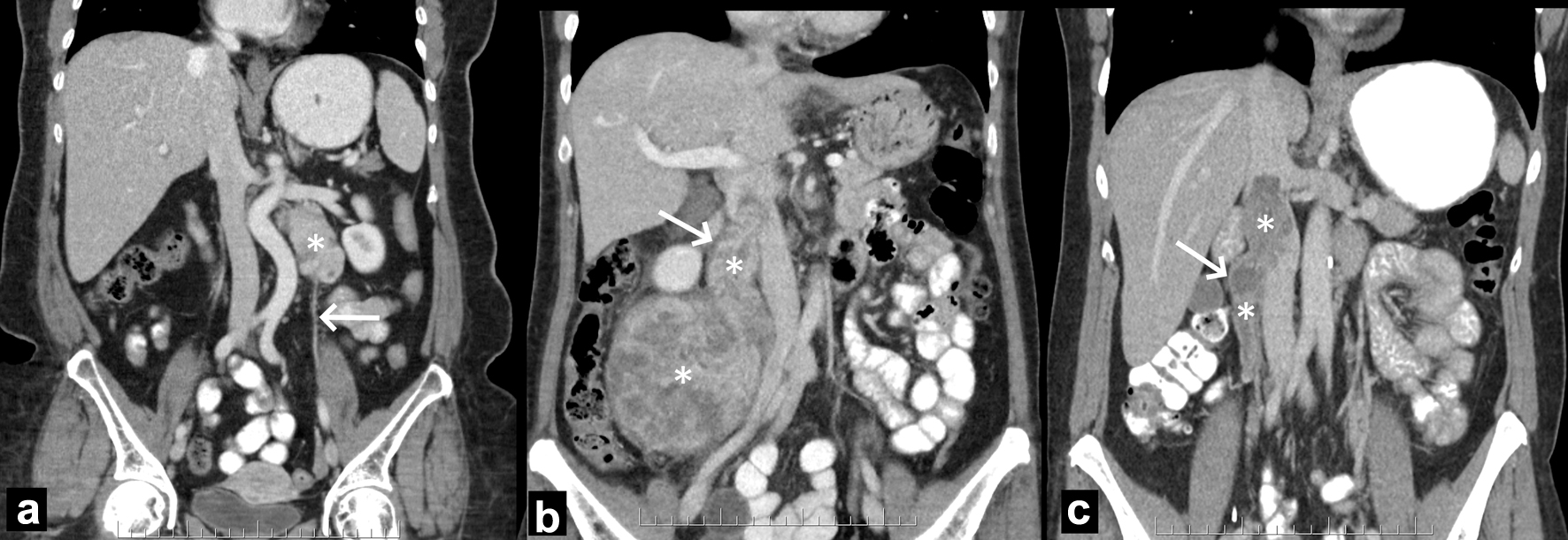
Figure 1. Illustration of designation of tumor origin in three different patients. (a) Left ovarian vein LMS. The left retroperitoneal tumor (asterisk) is located caudally to the left renal vein and laterally to the aorta. The left ovarian vein (arrow) extends into and from the tumor (not shown). (b) Right ovarian vein LMS. The right retroperitoneal tumor (asterisks) extends up the right ovarian vein (arrow) and into the IVC. (c) IVC IIA. The purely intravascular tumor (asterisks) lies within the right ovarian vein (arrow) and IVC, at and below the left renal vein. Because the tumor had a wider diameter in the IVC than in the ovarian vein, the former was designated as site of origin. IVC: inferior vena cava; LMS: leiomyosarcoma.
Figure 2.
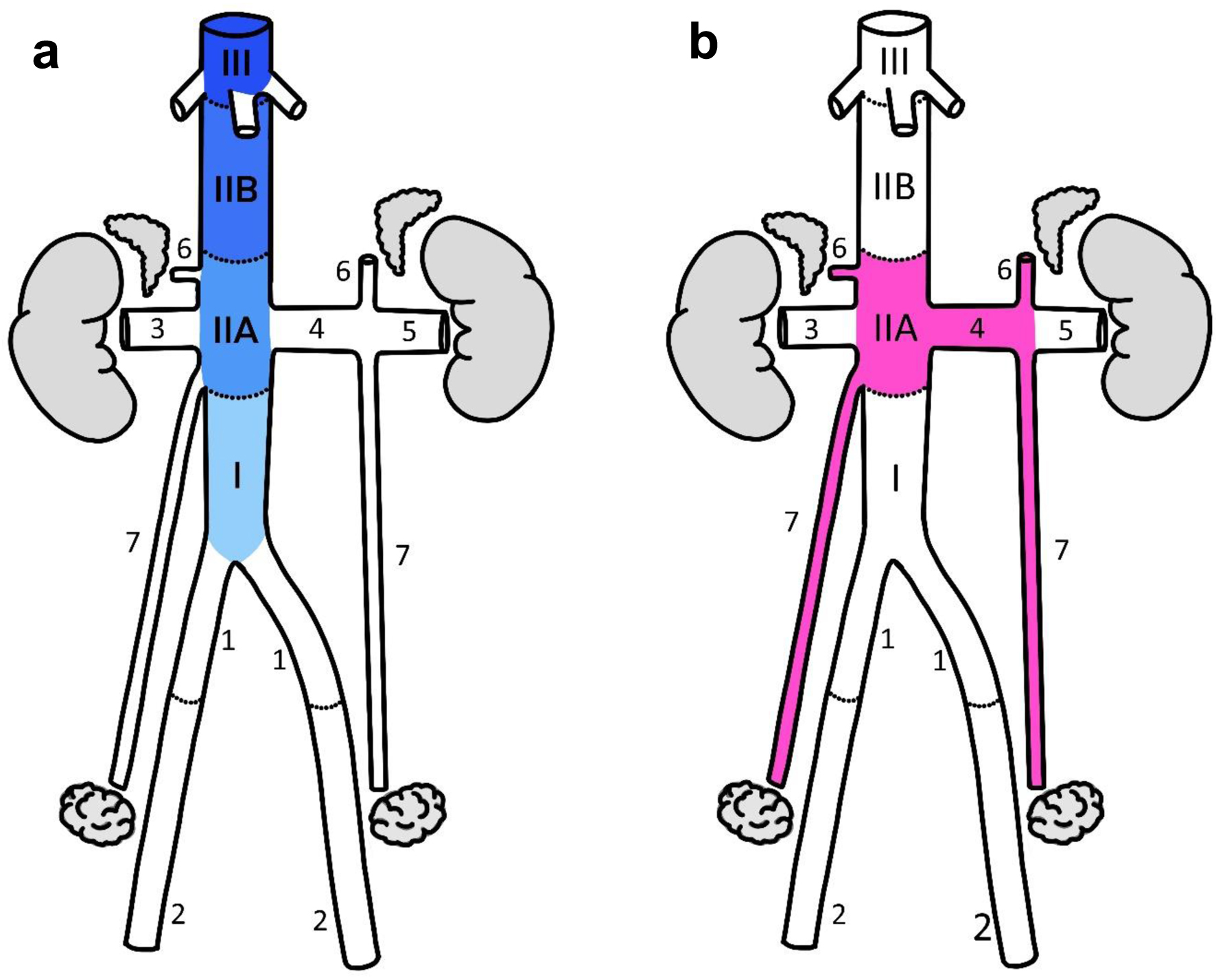
Figure 2. (a) IVC segments. The notation used is based on prior studies of IVC LMS with the following modifications: 1 - the cranial limit of IVC I was defined at the junction of the gonadal vein or either renal vein, whichever was more inferior; 2 - IVC II was divided into A and B segments with the hepatic margin as their boundary. (b) Sex-hormone drainage pathway (SHDP). The first and immediate second veins draining the hormone-producing organs were included as part of the drainage pathway. Anatomical annotations: I, IIA, IIB, III, IVC segments; 1 - common iliac vein, 2 - external iliac vein, 3 - right renal vein, 4 - medial left renal vein, 5 - lateral left renal vein, 6 - adrenal vein, 7 - ovarian vein. IVC: inferior vena cava; LMS: leiomyosarcoma.
Figure 3.
Figure 3. Breakdown of derivation of the final population.
Figure 4.
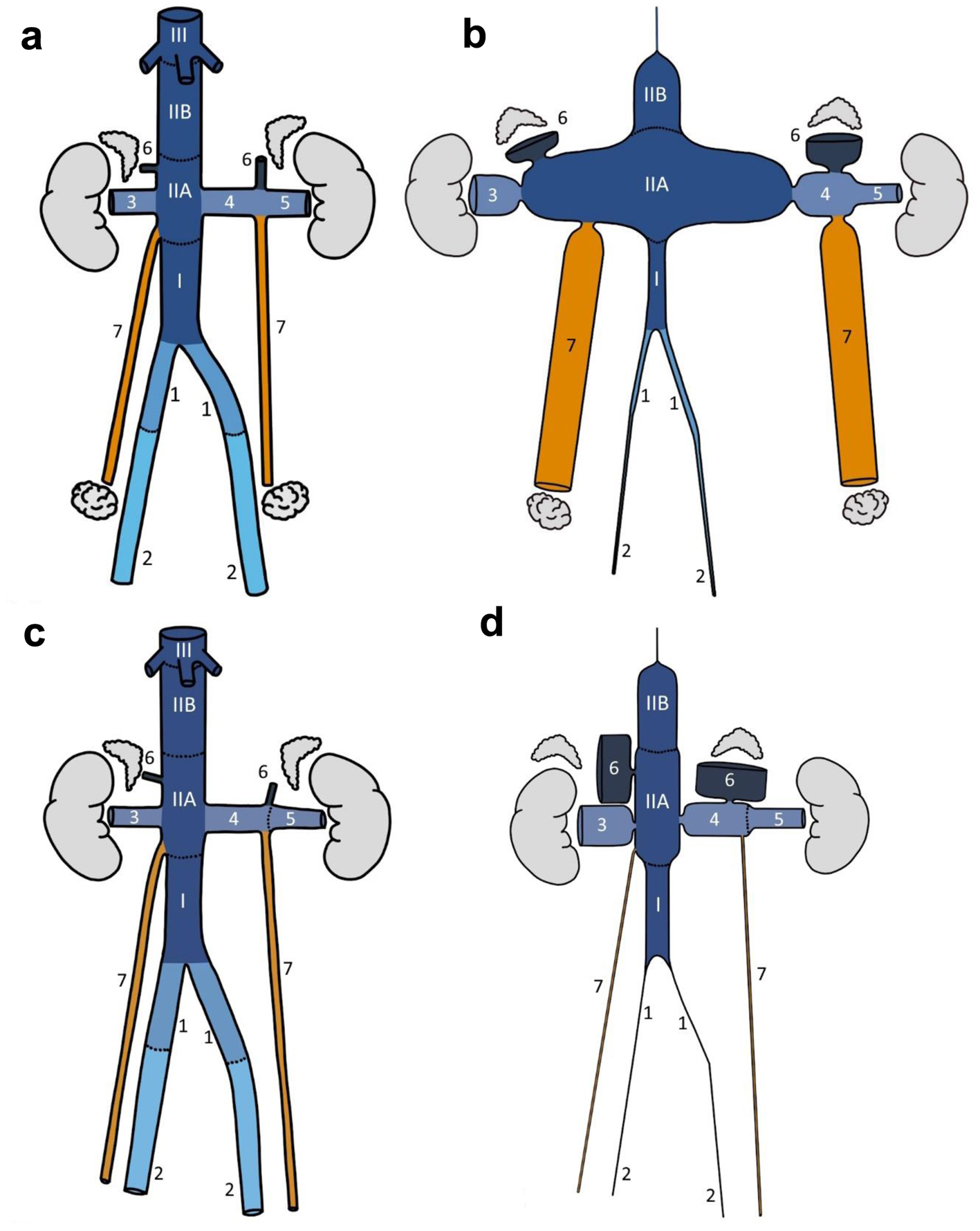
Figure 4. Normal anatomy (a: female; c: male) and proportional topographical (b: female; d: male) diagrams of selected retroperitoneal veins. The normal vessel lengths and diameter were drawn based on averages from 10 female/male patients in the study cohort. Proportional topographical diagrams are distortions of the anatomy normalized to one variable. For example, the cortical sensory homunculus is a proportional topographical diagram of the body distorted based on the size of the brain cortex supplying the various anatomical regions. These venous proportional topographical diagrams were obtained by altering the vessel diameter based on incidence per mm2 and then normalized to IVC IIB segment. All altered vessels are therefore proportional to IVC IIB segment. Anatomical annotations: I, IIA, IIB, III, IVC segments; 1 - common iliac vein, 2 - external iliac vein, 3 - right renal vein, 4 - medial left renal vein, 5 - lateral left renal vein, 6 - adrenal vein, 7 - ovarian/testicular vein.

Tables

Table 1. Patient Demographics
 
VariableAll patients, N (%)Females, N (%)Males, N (%)P
aOvarian (1), adrenal (3.5) and paratesticular (10) LMS. P values refer to differences between females and males. FNCLCC: French Federation of Cancer Centers Sarcoma Group; ER/PR: estrogen receptor/progesterone receptor; N/A: not applicable.
Number of patients15595 (61.3)60 (38.7)< 0.01
Age at diagnosis, years, median (range)60 (22 - 90)60 (22 - 90)61 (23 - 88)0.6
Tumour size, cm, median (range)7.8 (1.1 - 30.3)8.2 (1.4 - 30.0)7.7 (1.1 - 30.3)0.7
FNCLCC grade, n (%)0.9
  122 (14.2)14 (14.7)8 (13.3)
  262 (40.0)38 (40.0)24 (40.0)
  351 (32.9)30 (31.6)21 (35.0)
  Unknown20 (12.9)13 (13.7)7 (11.7)
Metastases at presentation, n (%)
  Absent132 (85.2)84 (88.4)48 (80.0)0.2
  Present23 (14.8)11 (11.6)12 (20.0)
Resection of primary, n (%)
  Yes124 (80.0)80 (84.2)44 (73.3)0.1
  No31 (20.0)15 (15.8)16 (26.7)
ER/PR positivity, n (%)
  Absent26 (16.8)24 (25.3)2 (3.3)0.6
  Present24 (15.5)21 (22.1)3 (5.0)
  Unknown105 (67.7)50 (52.6)55 (91.7)
Venous origin
  Yes89 (57.4)66 (69.5)23 (38.3)< 0.0001
  No66 (42.6)29 (30.5)37 (61.7)
Organ system, n (%)
  Sex-hormone drainage pathwaya82 (52.9)58 (61.1)24a (40.0)0.01
  Non-sex-steroid drainage pathway73 (47.1)37 (38.9)36 (60.0)
Ovarian drainage pathway, n (%)
  Yes53 (34.2)53 (55.8)0 (0.0)N/A
  No102 (65.8)42 (44.2)60 (100.0)


Table 2. Distribution of Tumor Location in Order of Frequency by Gender
 
LocationFemales (N = 95)LocationMales (N = 60)
n%n%
If the organ/vein of origin was narrowed to two sites, both sites were recorded but given half a point each. If more than two possible sites were noted, the location was deemed indeterminate. IVC: inferior vena cava.
VenousVenous
  Ovarian vein/ovary2728.4  Renal vein58.3
  IVC IIA21.522.6  IVC IIA46.7
  Renal vein66.3  Other veins3.55.8
  Adrenal/adrenal vein5.55.8  Adrenal/adrenal vein3.55.8
  IVC IIB55.3  IVC IIB35
  Anal/sphincter/perineum55.3  IVC I23.3
  IVC I33.2  Testicular vein11.7
  Other veins22.1  IVC III00
  IVC III00
Non-venousNon-venous
  Anal/sphincter/perineum55.3  Paratesticular/spermatic cord1016.7
  Indeterminate44.2  Prostate610
  Vagina33.2  Rectum46.7
  Colon33.2  Liver35
  Small bowel22.1  Other genitals35
  Stomach22.1  Indeterminate35
  Bladder22.1  Kidney2.54.2
  Rectum11.1  Bladder23.3
  Liver11.1  Other bowel23.3
  Kidney11.1  Stomach1.52.5
  Artery11.1  Ureter11.7
  Ureter00


Table 3. The Absolute and Normalized Rates of Venous AV-LMS by Sex
 
SexVein/vein segmentFrequency/mm2Normalized rate (IVC IIB = 1)
AV-LMS: abdominal venous leiomyosarcoma; IVC: inferior vena cava.
FemaleAdrenal veins1.3 × 10-214.3
IVC IIA5.8 × 10-36.2
Ovarian veins4.6 × 10-34.9
Right renal vein1.9 × 10-32.1
Left renal vein, medial1.7 × 10-31.8
Left renal vein, lateral9.4 × 10-41.0
IVC IIB9.4 × 10-41.0
IVC I6.9 × 10-40.7
Common iliac veins1.7 × 10-40.2
External iliac veins1.2 × 10-40.1
IVC III00
MaleAdrenal veins1.1 × 10-218.4
Right renal vein1.5 × 10-32.5
Left renal vein, medial8.9 × 10-41.5
Left renal vein, lateral7.6 × 10-41.3
IVC IIA7.0 × 10-41.2
IVC IIB6.1 × 10-41.0
IVC I4.0 × 10-40.7
Testicular veins1.7 × 10-40.3
Common iliac veins00
External iliac veins00
IVC III00


Table 4. Univariate and Multivariable Logistic Regression of Factors Associated With Metastasis at Presentation for the Entire Cohort (n = 155)
 
VariablesUnivariateMultivariable
OR95% CIPOR95% CIP
CI: confidence interval; FNCLCC: French Federation of Cancer Centers Sarcoma Group; OR: odds ratio.
Sex0.155
  Male--
  Female0.50.2 - 1.3
Age (years)1.01.0 - 1.00.960
Tumor size (cm)1.11.0 - 1.20.0031.91.2 - 3.20.009
Organ system0.0080.020
  Sex-hormone drainage pathway----
  Non-sex-hormone drainage pathway3.81.5 - 11.53.31.2 - 9.3
Ovarian drainage pathway--0.075
  Yes2.81.0 - 10.1
  No
FNCLCC grade0.308
  1--
  21.50.3 - 10.4
  32.80.7 - 18.9


Table 5. Univariate and Multivariable Logistic Regression of Factors Associated With Metastasis at Presentation for the Cohort With Exclusion of Paratesticular Tumors (n = 145)
 
VariablesUnivariateMultivariable
OR95% CIPOR95% CIP
CI: confidence interval; FNCLCC: French Federation of Cancer Centers Sarcoma Group; OR: odds ratio.
Sex0.10
  Male--
  Female0.50.2 - 1.2
Age (years)1.01.0 - 1.00.80
Tumor size (cm)1.11.0 - 1.20.0032.01.2 - 3.40.006
Organ system0.0090.016
  Sex-hormone drainage pathway----
  Non-sex-hormone drainage pathway4.11.4 - 12.03.81.3 - 11.2
Ovarian drainage pathway--0.06
  Yes3.01.0 - 9.4
  No
FNCLCC grade0.40
  1--
  21.70.4 - 11.7
  32.70.6 - 18.7