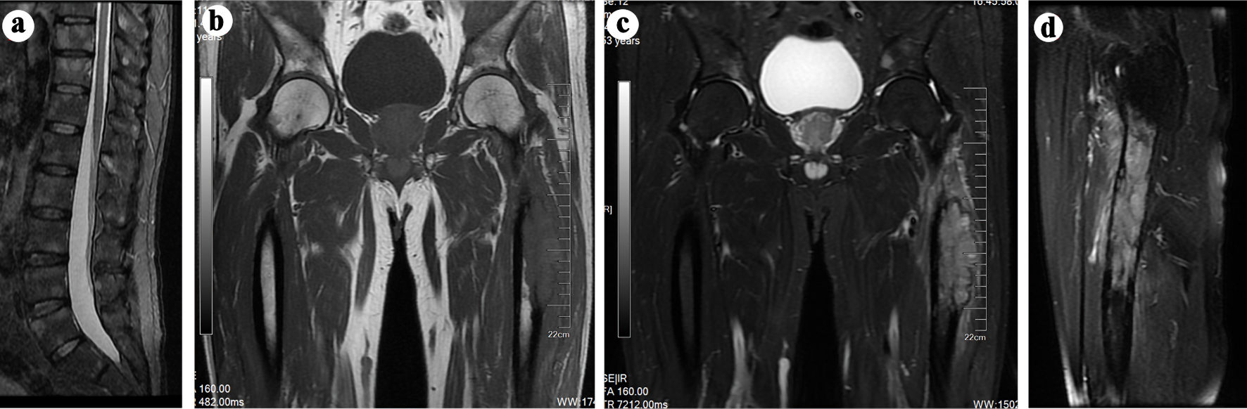
Figure 1. (a-c) MRI scans indicating potential multiple metastatic lesions in bilateral proximal femur, ilium, ischium, pubis, and spine. (b) T1-weighted sagittal spin-echo sequence of the thigh and hip joint displaying a hypointense lesion in the left proximal femur. (c, d) T2-weighted sagittal spin-echo sequence with fat-suppression demonstrating a hyperintense lesion involving intraosseous and extraosseous soft tissue in the left proximal femur. MRI: magnetic resonance imaging.
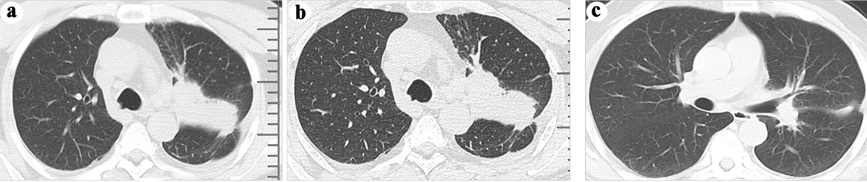
Figure 2. (a) A neoplasm in the left upper lobe was observed on a computed tomography (CT) scan. (b) The presence of a neoplasm in the left lung was confirmed through an enhanced CT scan. (c) A follow-up CT scan at 3 months after osimertinib administration revealed shrinkage of the neoplasm in the left upper lobe.
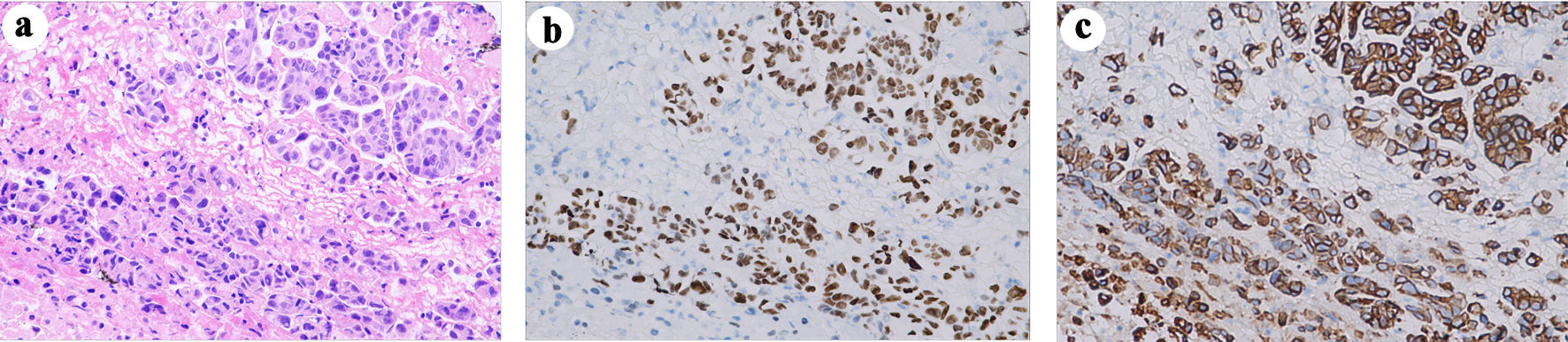
Figure 3. (a) H&E staining (original magnification, × 400) depicting tumor cell morphology. Immunohistochemical analysis revealed expressions of CK7 ((b) original magnification, × 400) and TTF1 ((c) original magnification, × 400), confirming the diagnosis of lung adenocarcinoma. H&E: hematoxylin and eosin; CK7: cytokeratin 7; TTF1: thyroid transcription factor 1.