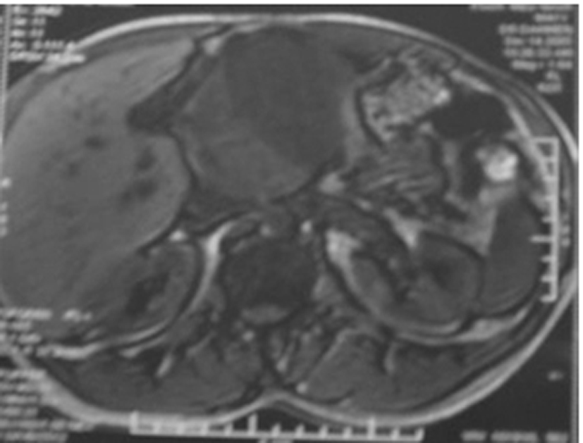
Figure 1. Magnetic resonance imaging: large retroperitoneal mass compressing the liver.
| World Journal of Oncology, ISSN 1920-4531 print, 1920-454X online, Open Access |
| Article copyright, the authors; Journal compilation copyright, World J Oncol and Elmer Press Inc |
| Journal website http://www.wjon.org |
Case Report
Volume 1, Number 2, April 2010, pages 94-96
Retroperitoneal Inflammatory Myofibroblastic Tumor: Case Report and Immunohistochemistry Study
Figures