World Journal of Oncology, ISSN 1920-4531 print, 1920-454X online, Open Access
Article copyright, the authors; Journal compilation copyright, World J Oncol and Elmer Press Inc
Journal website http://www.wjon.org

Case Report

Volume 1, Number 4, August 2010, pages 179-181


Peritoneal Malignant Psammomatous Mesothelioma

Figures

Figure 1.
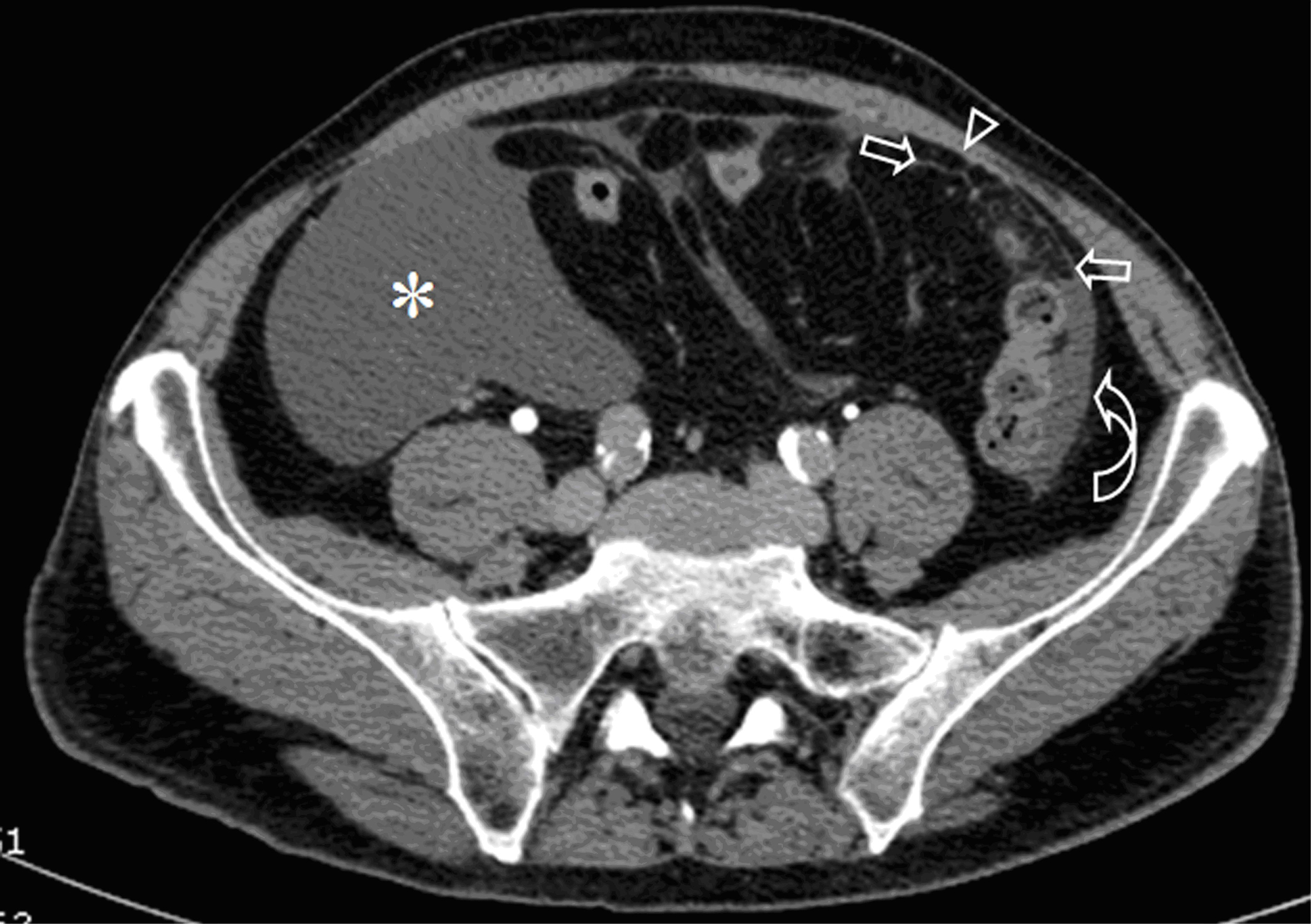
Figure 1. Contrast-enhanced CT of the abdomen shows abundant peritoneal fluid in the right iliac fossa (*) and paracolic fluid in the left side (curved arrow). Note the thick septa with micronodules in the greater omentum (arrows) and circumscribed enhancement of the thickened peritoneum (arrow head).
Figure 2.
Figure 2. A. The peritoneal bioptic fragment shows massive deposition of Psammoma bodies (H&E; 25X). B. The superficial peritoneum shows a proliferation of malignant cells with massive presence of psammoma bodies (H&E; 100X).
Figure 3.
Figure 3. Neoplastic cells show nuclear and cytoplasmic calretinin immunoreactivity.
Figure 4.
Figure 4. A positive membranous staining with the D2-40 antibody is seen in neoplastic cells.