Figures
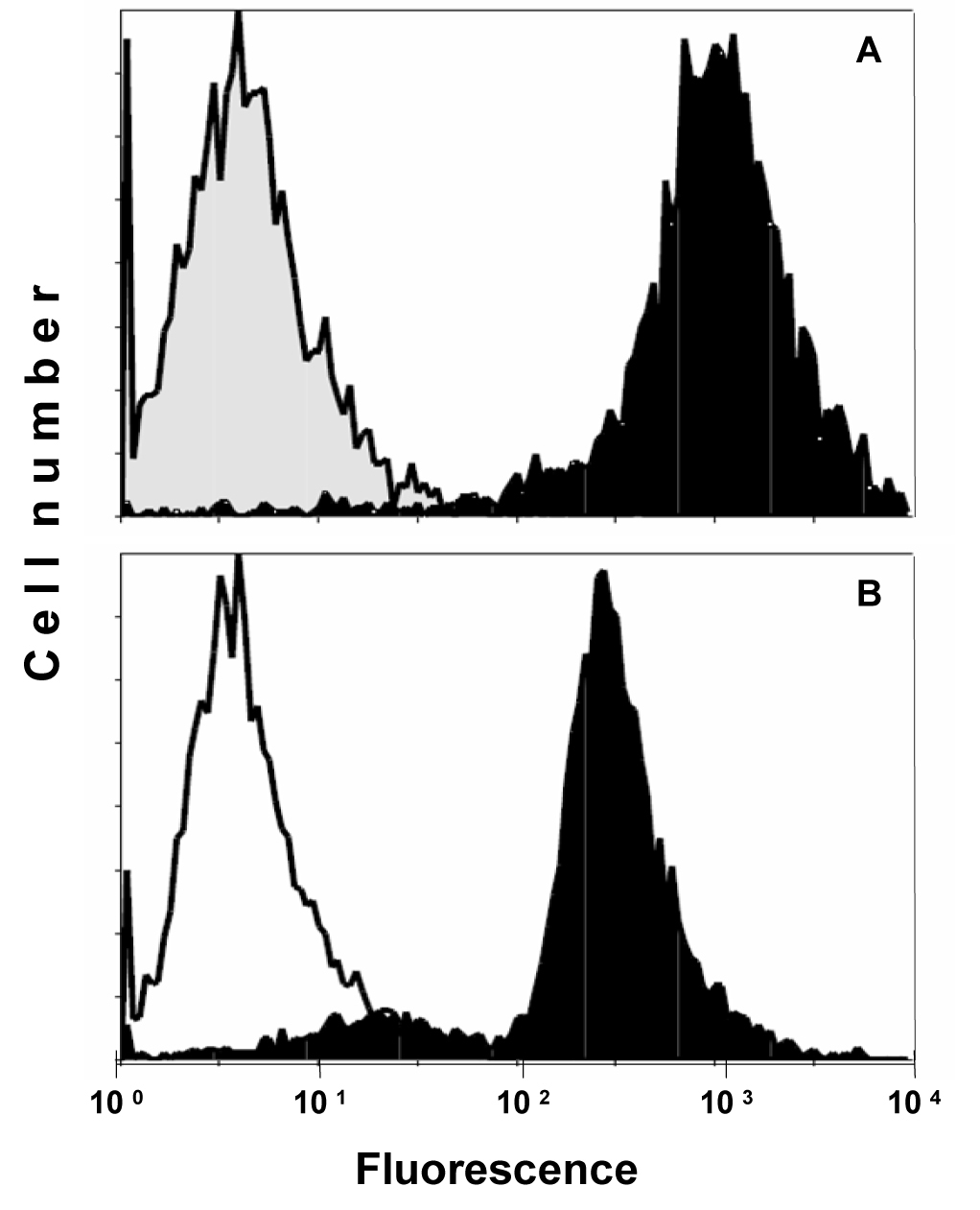
Figure 1. Flow cytometric analysis of CD99/MIC2 HBA-71 antigen expression of (A) KAL EFT cells and (B) AHTO-7 osteoblasts. Immunofluorescence of the cells stained with HBA-71 is shown in black, the NIC-1 isotype controls in grey (KAL) and white (AHTO-7), respectively.
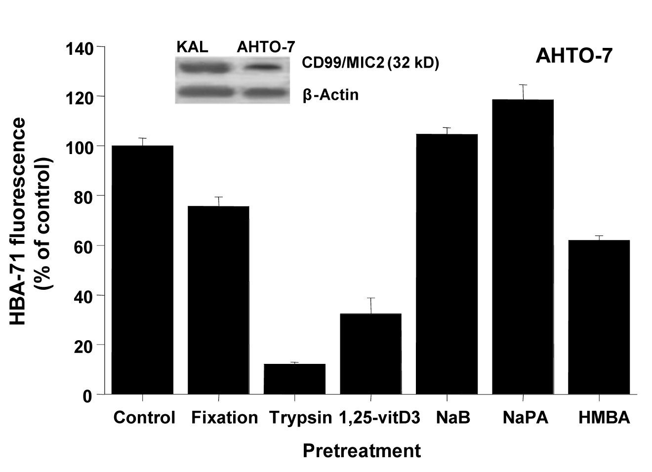
Figure 2. CD99/MIC2 Western blot and effects of fixation, trypsin treatment and differentiation inducers on HBA-71 expression of AHTO-7 osteoblasts. The CD99/MIC2 antigen was blotted with the HBA-71 antibody using extracts of KAL and AHTO-7 cells, respectively. Protein amounts of β-actin are shown as controls. Relative fluorescence intensities are expressed as mean ± SD (n = 3). With the exception of NaB pretreatment, all differences to the control are statistically significant.
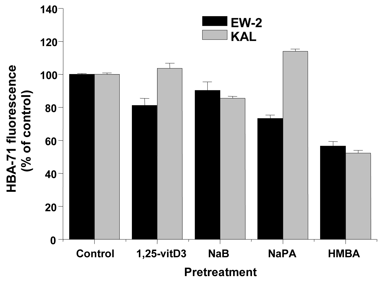
Figure 3. Effects of differentiation inducers on HBA-71 immunofluorescence of EW-2 and KAL EFT cells. Relative fluorescence intensities are expressed as mean ± SD (n = 3). All differences to the respective controls are statistically significant.
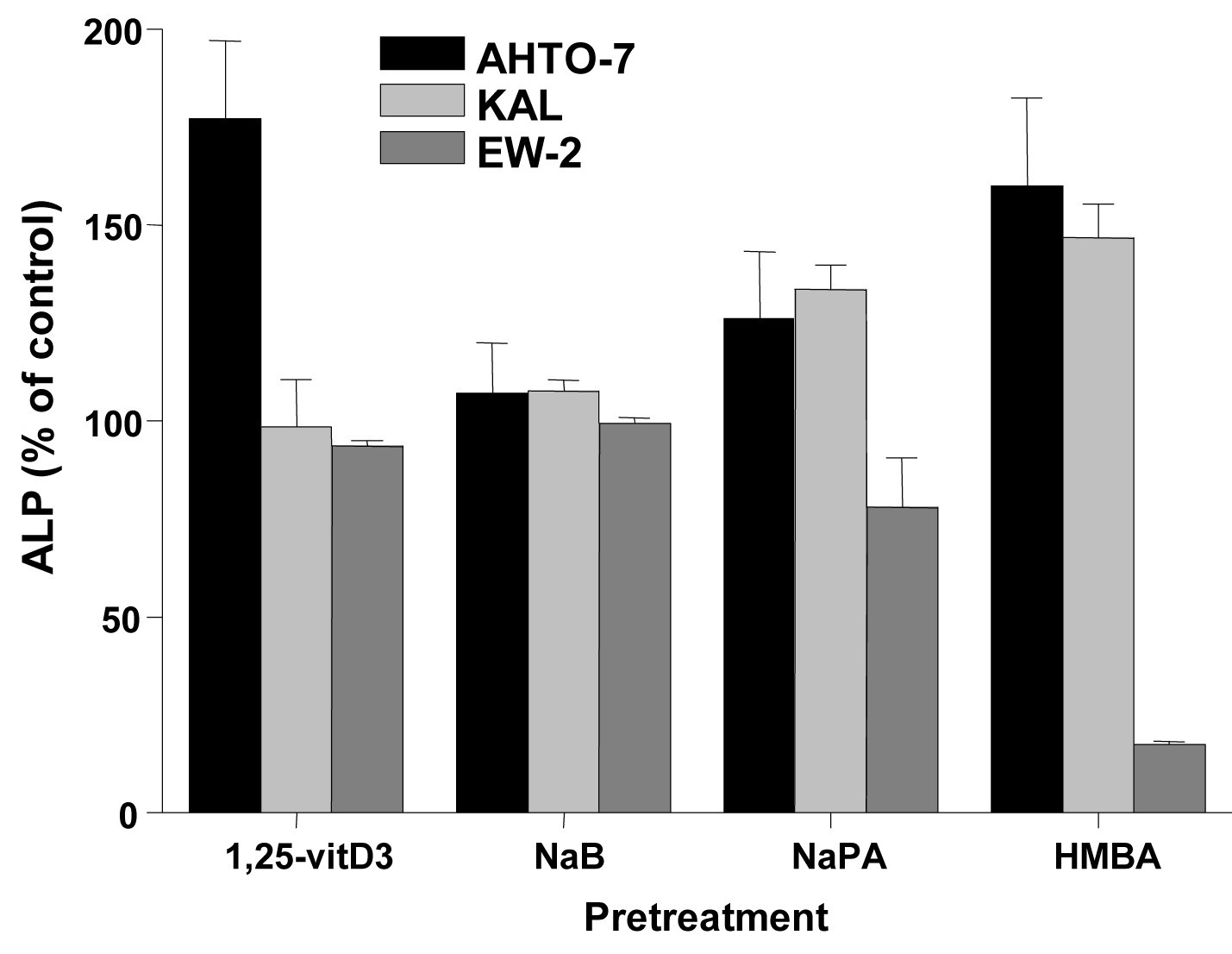
Figure 4. Changes in cellular ALP activity in response to differentiation inducers of AHTO-7, KAL and EW-2 cells. Activity of ALP is expressed as mean ± SD (n = 3). All differences to the control are statistically significant, except for 1, 25-vitD3 and NaB treatment of EW-2 cells.