World Journal of Oncology, ISSN 1920-4531 print, 1920-454X online, Open Access
Article copyright, the authors; Journal compilation copyright, World J Oncol and Elmer Press Inc
Journal website http://www.wjon.org

Case Report

Volume 3, Number 3, June 2012, pages 119-123


A Case of Angiosarcoma Arising in Trunk of the Right Pulmonary Artery Clinically Simulating Pulmonary Thromboembolism

Figures

Figure 1.
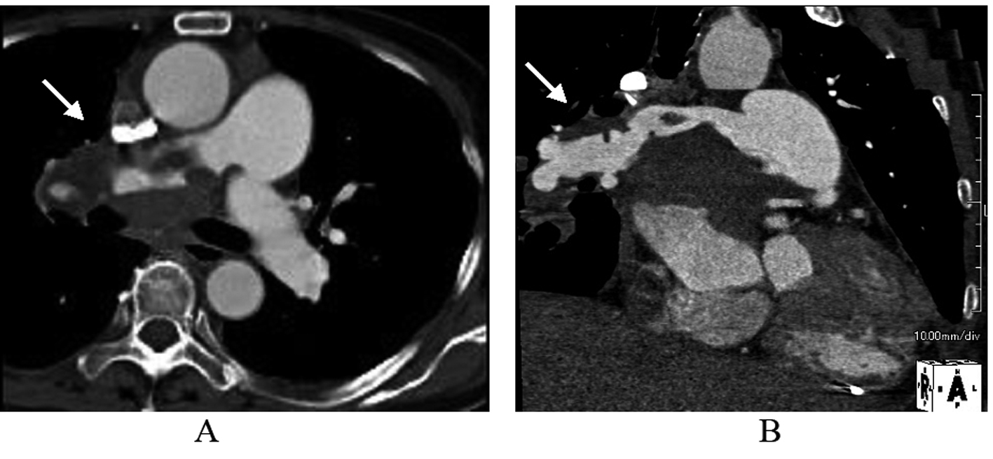
Figure 1. The image of Contrast-enhanced computed tomography scan. Early arterial phase showed massive filling defects in the proximal portion of right pulmonary artery and around the left atrium in axial (A) and sagittal (B) views.
Figure 2.
Figure 2. The image of right pulmonary angiography. Vascularized mass (white arrow) with a fistula from the right pulmonary artery to the left atrium (black arrow).
Figure 3.
Figure 3. The image of maximum intensity projection image of 18-fluorodeoxyglucose positron emission tomography (FDG-PET). Increased uptake of FDG at the area of the right pulmonary artery (white arrow) and the bilateral atria (yellow arrow) in coronal (A) and axial (B) views.
Figure 4.
Figure 4. A. Macroscopical finding showed that the right main trunk and its second branched segments of the pulmonary artery were completely occluded by the tumor and thrombus. A red arrow indicates the right pulmonary artery. B. Hematoxilin and eosin staining showed that the tumor cells were composed of pleomorphic cells which had large and vesicular nuclei with prominent nucleori and abundant eosinophilic cytoplasm. C. Elastica and masson staining showed the destruction of the wall of the right pulmonary artery by the tumor cells. ‘Wall’ indicates the wall of the right main trunk of the pulmonary artery, and ‘Lumen’ indicates its lumen. D. D, E and F: Immunohistochemical findings showed the tumor cells were diffusely positive for vimentin (D), focally positive for CD31 (E) and CD34 (F) in the vascular channel area.