World Journal of Oncology, ISSN 1920-4531 print, 1920-454X online, Open Access
Article copyright, the authors; Journal compilation copyright, World J Oncol and Elmer Press Inc
Journal website http://www.wjon.org

Case Report

Volume 3, Number 3, June 2012, pages 142-145


Superior and Inferior Extension of Carotid Body Tumors

Figures

Figure 1.
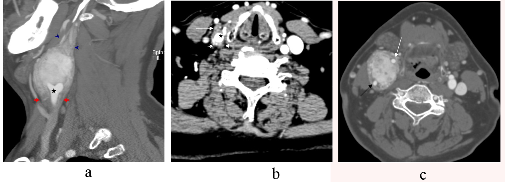
Figure 1. a-c. CT Angiography sagittal section showing a large carotid body tumor splaying the external carotid artery (ECA) and the internal carotid artery (ICA) at the level of th common carotid bifurcation (star). The tumor notably extends inferiorly 1.6 cm below the common carotid bifurcation (arrows). It also extends approximately 1.6 cm superiorly above the main bulk of the tumor (arrowhead) (a). Axial CT Angiography of the neck showing the tumor (long arrows) wrapping the common carotid artery (star) below the bifurcation with avid vascularity of the tumor (short arrows) (b). Axial CT Angiography section demonstrating an avidly enhancing mass splaying the internal carotid artery (black arrow) and the external carotid artery (white arrow) (c).
Figure 2.
Figure 2. a-d. B-mode sonography showing a solid, well-defined, and hypoechoic tumor splaying the internal carotid artery(ICA) and external carotid artery (ECA) (a). Color flow Doppler sonography demonstrates a large mass at the carotid bifurcation. There is splaying of the internal carotid artery (ICA) and the external carotid artery (ECA) starting from the level of the common carotid bifurcation(star) (b, c) with high vascularity (d).