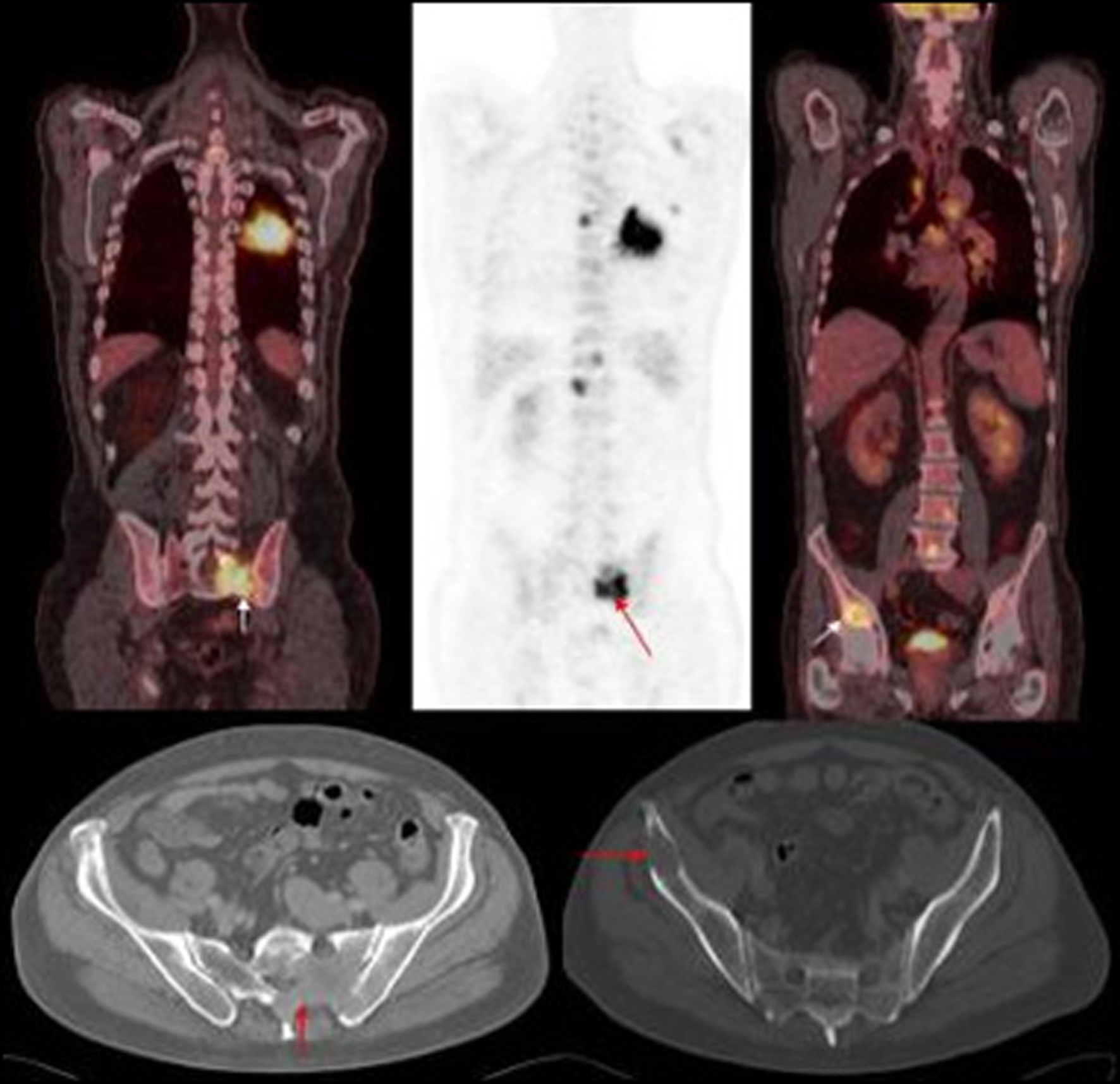
Figure 1. PET images of primary left lung cancer metastatic deposits in the bony pelvis and lumbar vertebrae with corresponding axial CT images of the pelvis.
| World Journal of Oncology, ISSN 1920-4531 print, 1920-454X online, Open Access |
| Article copyright, the authors; Journal compilation copyright, World J Oncol and Elmer Press Inc |
| Journal website http://www.wjon.org |
Original Article
Volume 3, Number 2, April 2012, pages 54-58
Is There a Role of Double Reporting and CT Pelvis for Lung Cancer Staging?
Figures
Table
| Infield | 21 | Outfield | 19 |
|---|---|---|---|
| Liver | 3 | Ilium | 5 |
| Adrenal | 5 | Pelvis | 4 |
| Ribs | 4 | Acetabulum | 2 |
| Scapula | 1 | L4 | 1 |
| Spine | 1 | High cervical nodes | 1 |
| Lung | 2 | Sacrum | 1 |
| Sternum | 1 | Proximal femur | 2 |
| Soft tissue | 2 | Glutei | 2 |
| Spleen | 1 | Post thigh | 1 |
| Subdiaphragmatic node | 2 |