World Journal of Oncology, ISSN 1920-4531 print, 1920-454X online, Open Access
Article copyright, the authors; Journal compilation copyright, World J Oncol and Elmer Press Inc
Journal website http://www.wjon.org

Case Report

Volume 4, Number 1, February 2013, pages 50-53


Papillary Renal Cell Carcinoma Presented With Supraclavicular Lymph Node Metastasis Without Renal Primary Lesion

Figures

Figure 1.
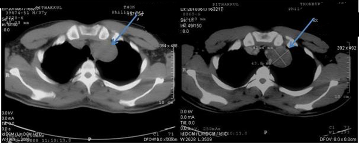
Figure 1. Pretreatment CT scan of chest in October 2008 (Pre-contrast on left side and post-contrast on right side) showing lobulated heterogeneous soft tissue density lesion at the apex of left lung.
Figure 2.
Figure 2. Pretreatment CT scan of abdomen in October 2008 (Pre-contrast on left side and post-contrast on right side) showing paraaortic lymph nodes enlargement on left side partially abuts wall of aorta and body of pancreas.
Figure 3.
Figure 3. Pretreatment CT scan of abdomen in October 2008 (Pre-contrast on left side and post-contrast on right side) showing enlargement of multilobulated left paraaortic node at the renal level, 7.4 cm in size. The mass displaces the left renal vein anteriorly. Some mass effect is noted with mildly dilated left renal pelvis.
Figure 4.
Figure 4. Hematoxylin & Eosin stain (H&E stain) of biopsied left supraclavicular lymph node showing lymph node with metastatic epithelial neoplasm arranging in papillary growth pattern. The neoplastic cells have enlarged pleomorphic and prominent nuclei, eosinophilic and clear neoplasm.