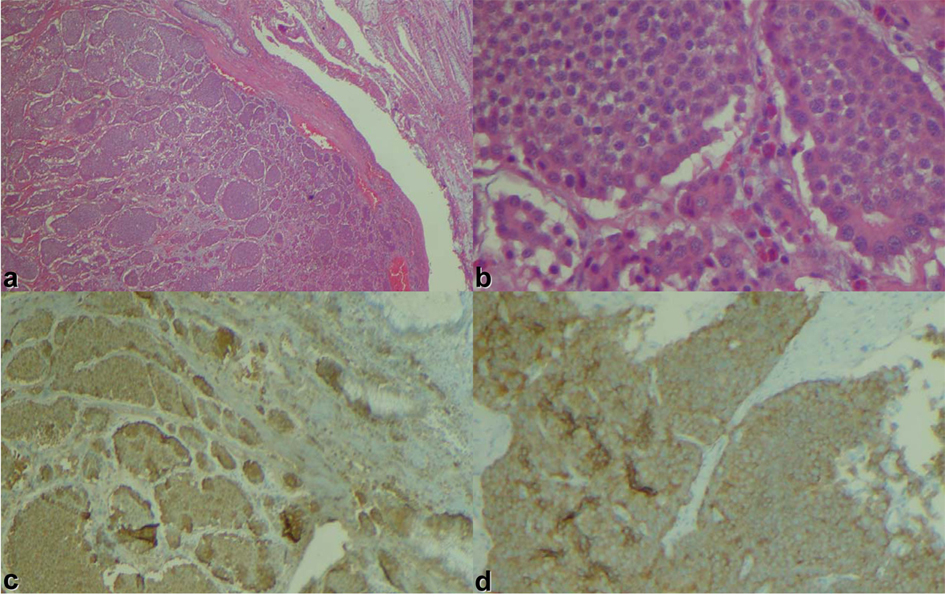
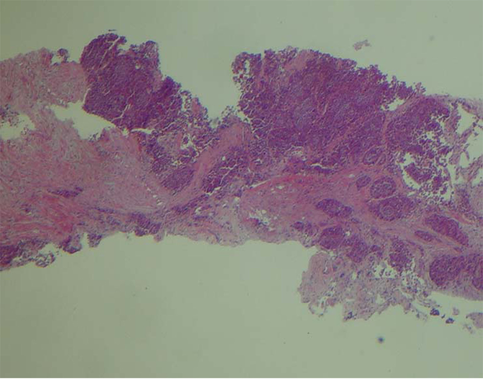
Figure 1. Small intestinal tumor in (a) low power view, (b) high power view, (c) chromogranin stain, (d) synaptophysin stain.
| World Journal of Oncology, ISSN 1920-4531 print, 1920-454X online, Open Access |
| Article copyright, the authors; Journal compilation copyright, World J Oncol and Elmer Press Inc |
| Journal website http://www.wjon.org |
Case Report
Volume 4, Number 2, April 2013, pages 114-117
Skeletal Muscle Metastases in a Patient With Neuroendocrine Tumor
Figures
Table
| Case 1 Quan GM, et al [9] | Case 2 Tiktinsky E, et al [10] | Case 3 Caobelli F, et al [11] | Our case | |
|---|---|---|---|---|
| Age/Sex | 53/M | 43/M | 70/M | 66/M |
| 1° site | Ileocecal | Lung | Ileal | Ileocecal |
| Size of 1° tumor | Not reported | Not reported | Not reported | 3.4 cm |
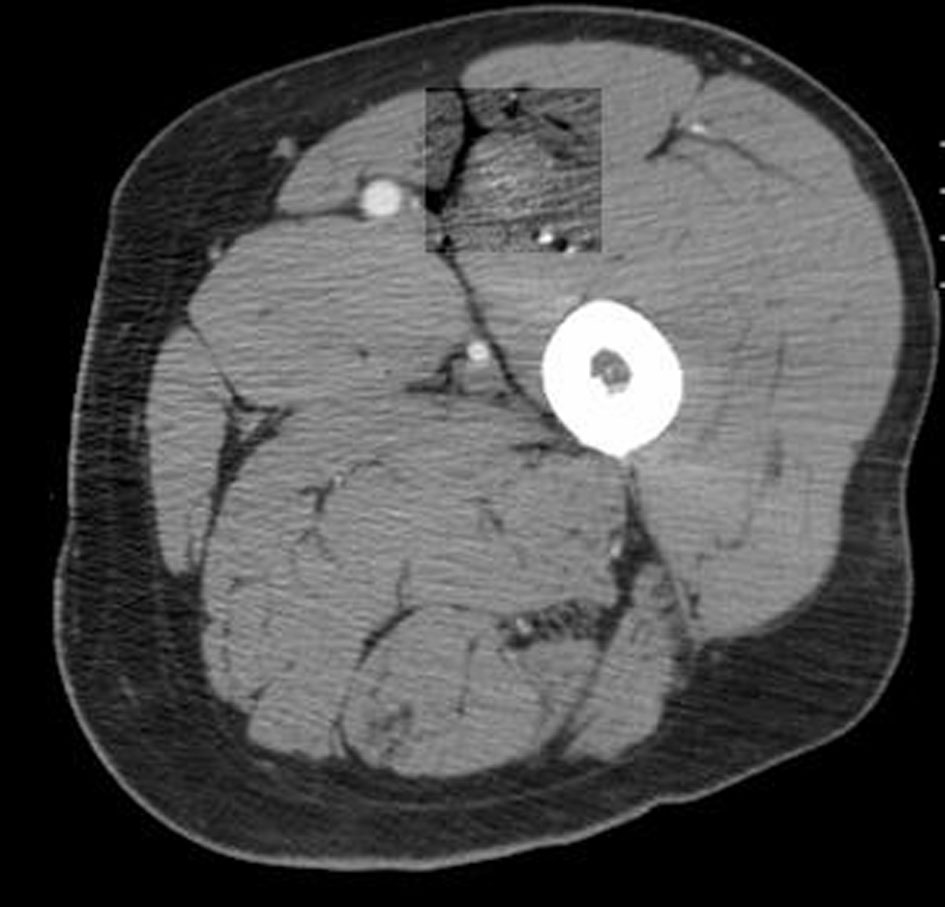
| Soft tissue mets | Popliteus muscle | Transverse abdominal muscle | Thigh muscle | Vastus intermedius |
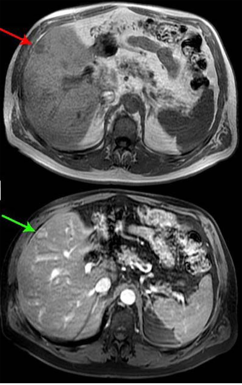
| Other site of metastasis | Liver | None | None | Liver |
| Time between initial diagnosis and presentation | 2.5 years | 2 months | 5 years | 6 months |
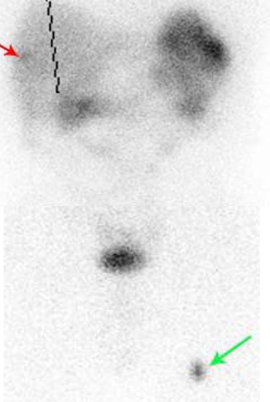
| Octreoscan | (+) | (+) | (+) | (+) |
| Biopsy | Metastatic malignant carcinoid | Atypical carcinoid | Carcinoid | Well differentiated neuroendocrine tumor |
| Treatment | Not reported | Not reported | Not reported | Octreotide LAR |