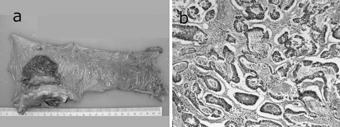
Figure 1. Resected specimen and pathological findings. a): Macroscopic appearance of surgically resected specimen showing type 2 advanced rectal cancer; b): Pathological examination showed a well-differentiated adenocarcinoma invading the perirectal tissues, with metastatic involvement in 1 of the 15 resected lymph nodes. (hematoxylin and eosin stain).

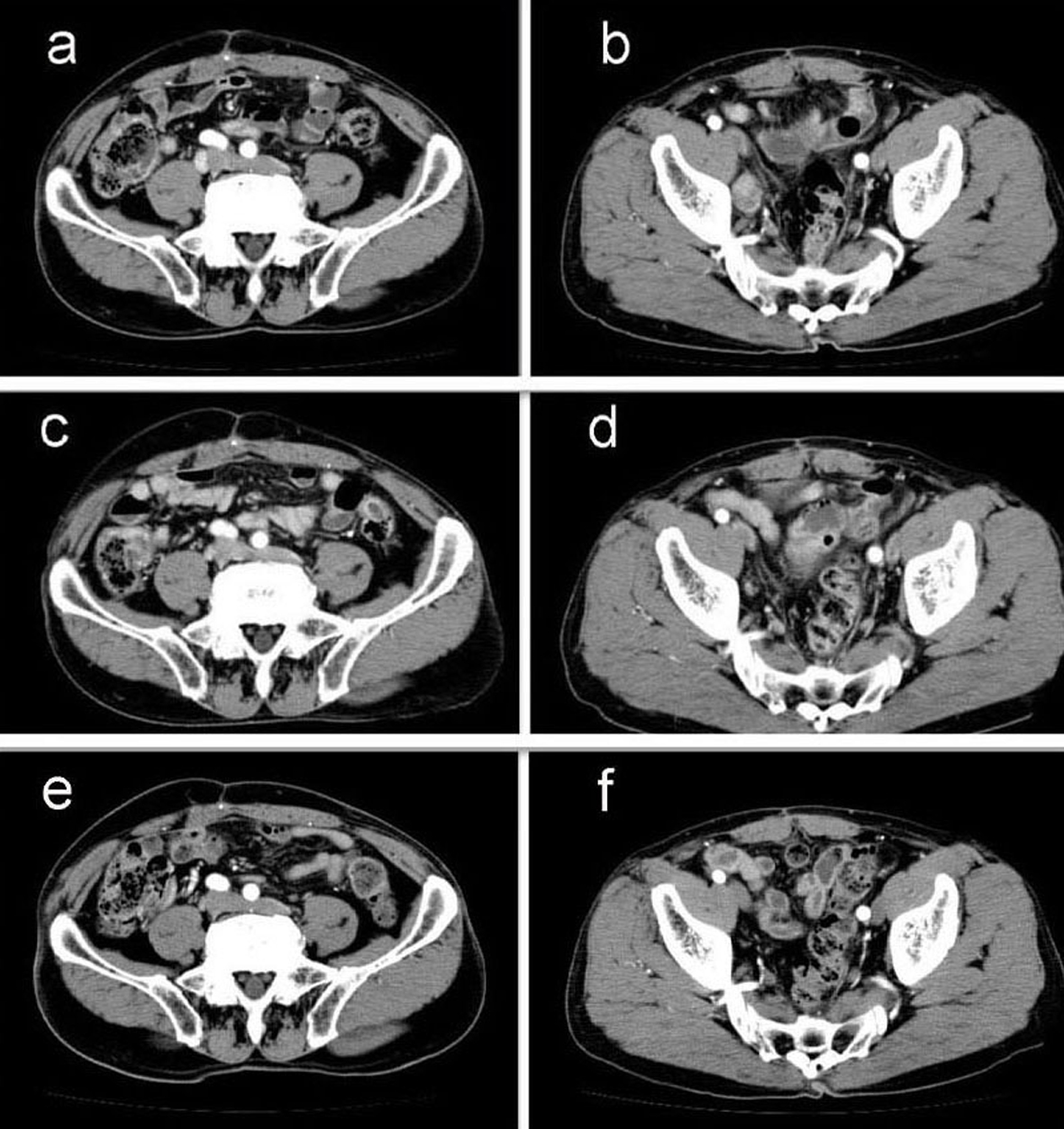
Figure 2. Abdominal computed tomography (CT) findings. a-b): CT scan before chemotherapy showing swollen para-aortic and right lateral pelvic lymph nodes; c-d): CT scan after 5 cycles of S-1 administration showing reduction in the swollen para-aortic lymph nodes; e-f): CT scan after radiation and 10 cycles of S-1 administration showing complete disappearance of swollen para-aortic and lateral pelvic lymph nodes. Hence, response in this case was classified as a complete response.

