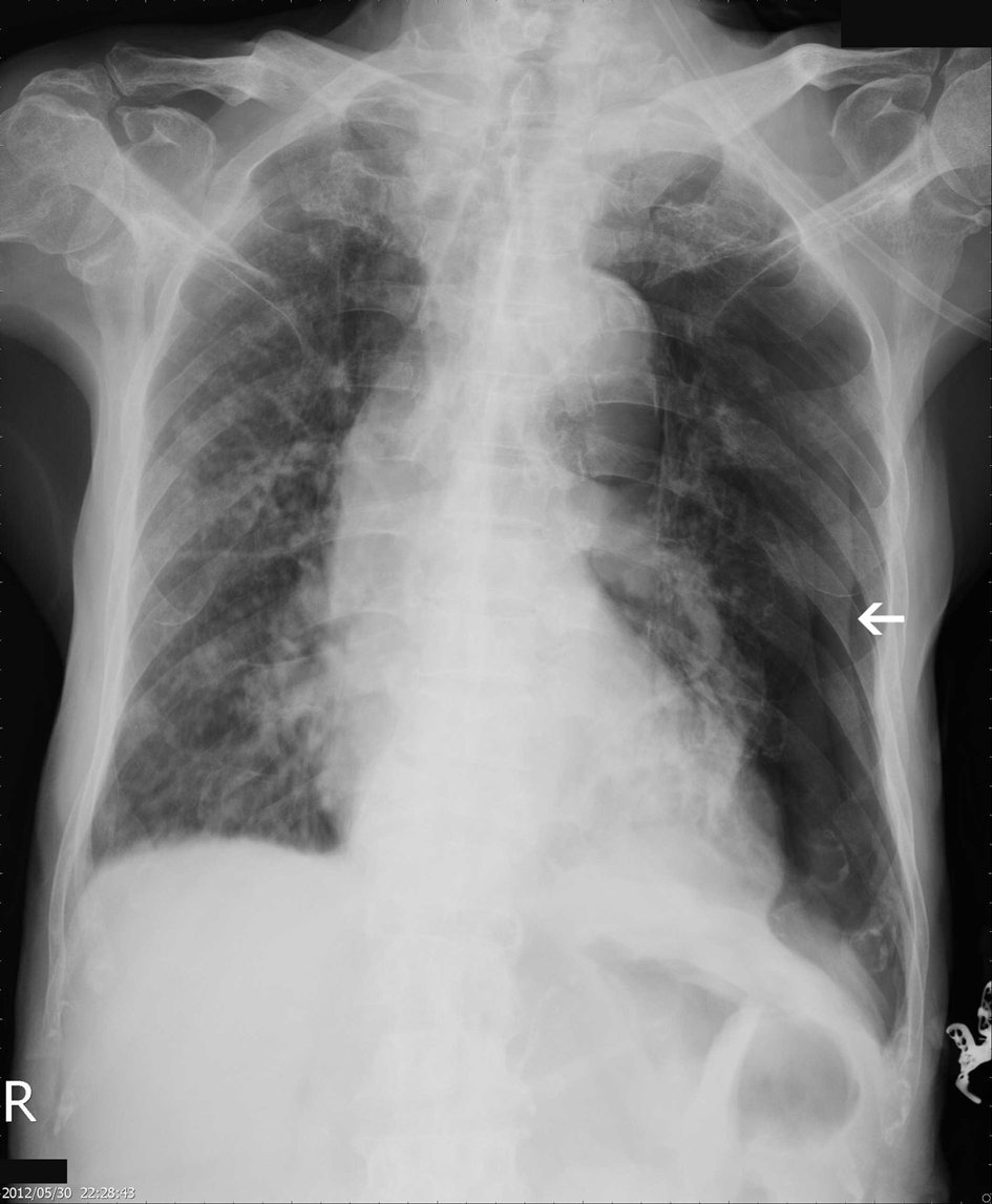
Figure 1. Initial CXR showing left pneumothorax, with ground grass densities over right lung.
| World Journal of Oncology, ISSN 1920-4531 print, 1920-454X online, Open Access |
| Article copyright, the authors; Journal compilation copyright, World J Oncol and Elmer Press Inc |
| Journal website http://www.wjon.org |
Case Report
Volume 4, Number 2, April 2013, pages 118-121
Pneumothorax as a Presenting Clinical Manifestation of Metastatic Prostate Cancer
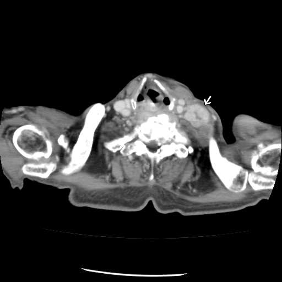
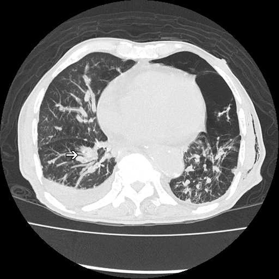
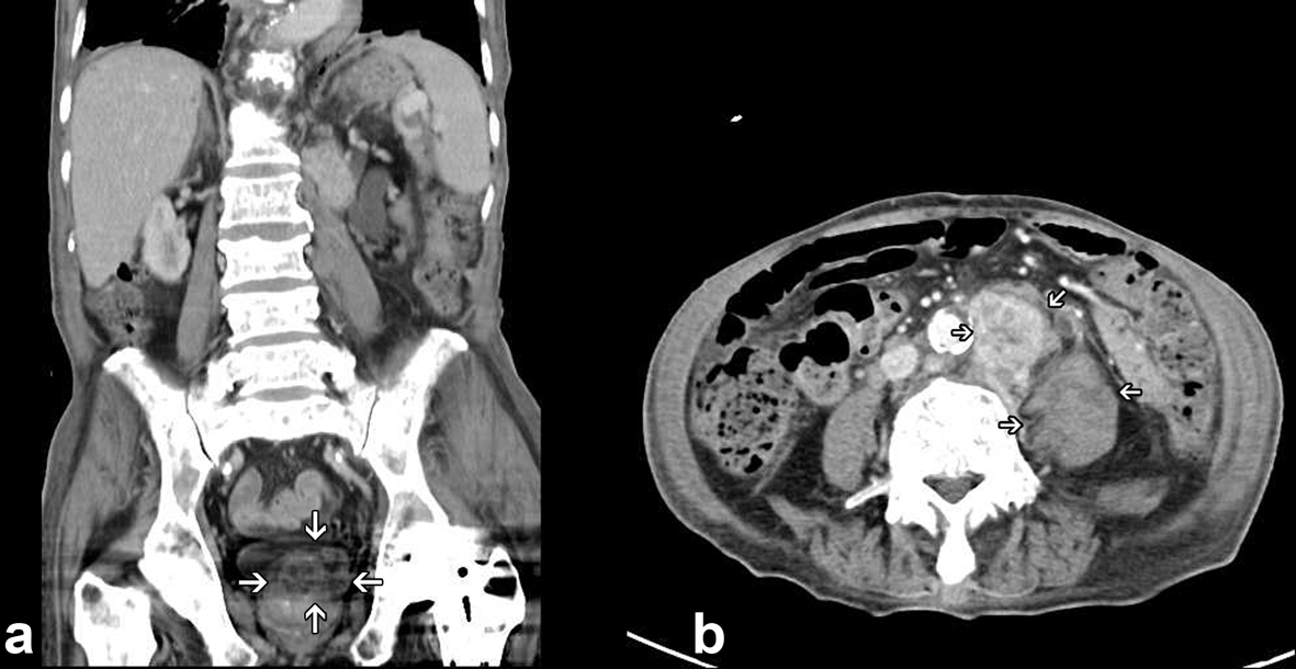
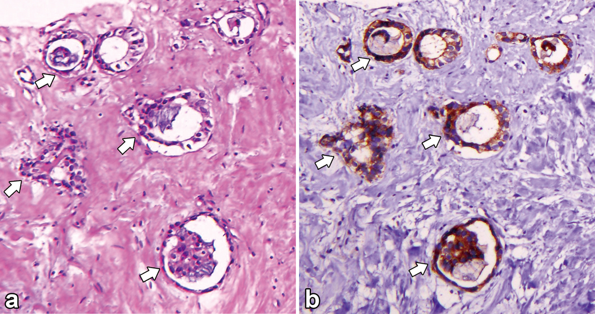
Figures