World Journal of Oncology, ISSN 1920-4531 print, 1920-454X online, Open Access
Article copyright, the authors; Journal compilation copyright, World J Oncol and Elmer Press Inc
Journal website http://www.wjon.org

Case Report

Volume 4, Number 4-5, October 2013, pages 210-213


Excellent Response to Palliative Chemotherapy for Pleural Recurrence of Uterine Papillary Serous Carcinoma

Figures

Figure 1.
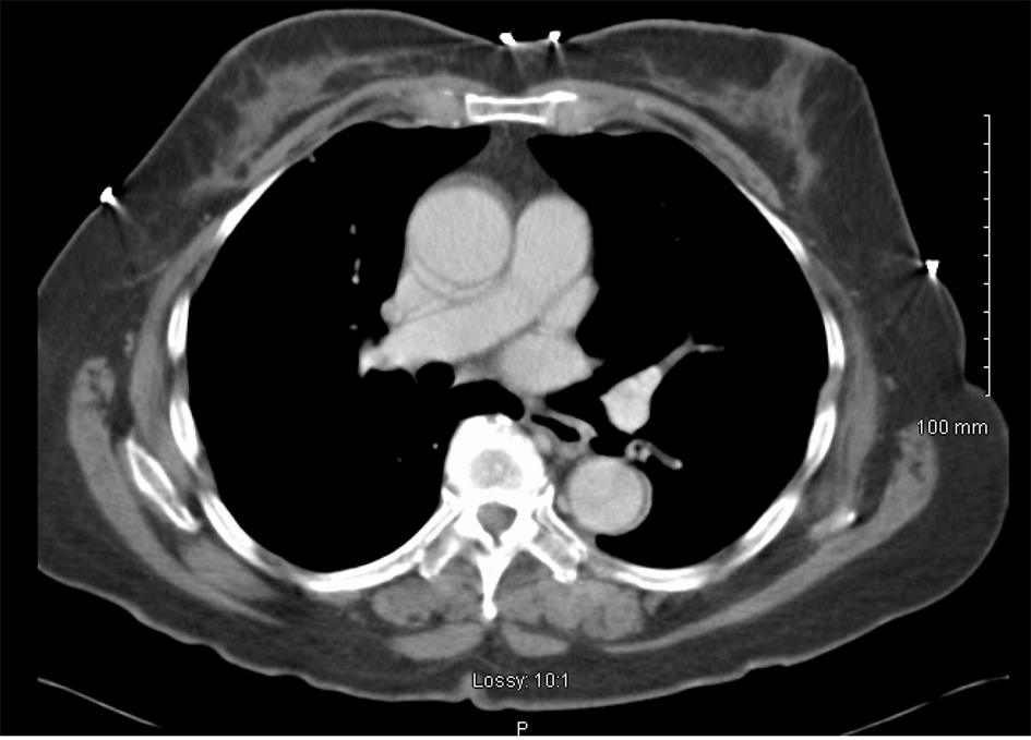
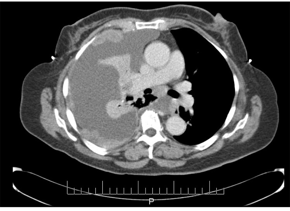
Figure 1. CT scan image demonstrating pleural effusion and nodularity.
Figure 2.
Figure 2. Pleural fluid Thin Prep demonstrating (A) Clusters of malignant epithelial cells in the background of necrotic cellular debris, 20 × (B) High power showing adenocarcinoma cells 100 × (C) 3-Dimensional clusters of adenocaricoma cells 100 ×
Figure 3.
Figure 3. Pleural fluid cell block demonstrating (A) Papillary clusters of adenocarcinoma on H&E 20 ×. (B) Arrow showing Psammoma body on H&E 100 ×.
Figure 4.
Figure 4. Pleural fluid cell block demonstrating (A) Immunostain positive for CK-7 (B) Immunostain positive for p53. (C) Immunostain negative for CK20.
Figure 5.
Figure 5. CT scan image after treatment demonstrating complete resolution of pleural effusion and nodularity.