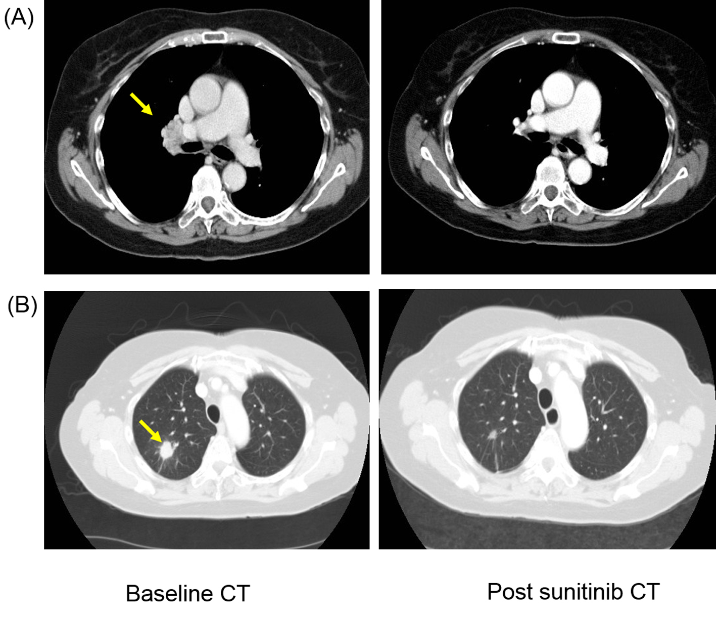
Figure 1. Patient developed response after first-line sunitinib therapy with complete resolution of (A) hilar lymphadenopathy (yellow arrow) and more than 50% reduction in size of (B) lung metastasis (yellow arrow).
| World Journal of Oncology, ISSN 1920-4531 print, 1920-454X online, Open Access |
| Article copyright, the authors; Journal compilation copyright, World J Oncol and Elmer Press Inc |
| Journal website http://www.wjon.org |
Case Report
Volume 5, Number 5-6, December 2014, pages 223-227
Pazopanib-Induced Regression of Brain Metastasis After Whole Brain Palliative Radiotherapy in Metastatic Renal Cell Cancer Progressing on First-Line Sunitinib: A Case Report
Figures