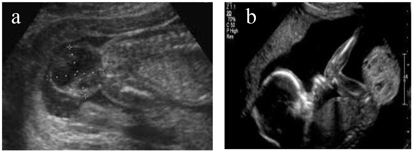
Figure 1. (a, b) Ultrasonography examination that showed mixed, solid/cystic tumorous oval mass in the sacral region of the embryo.
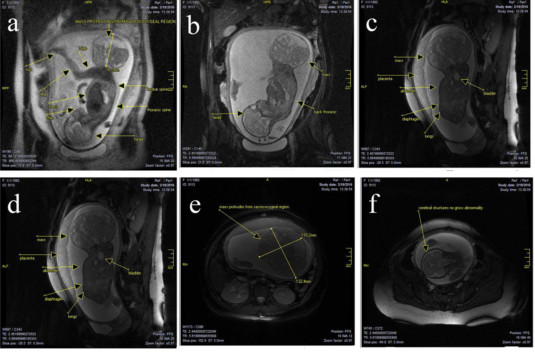
Figure 2. (a-f) There was a large well-circumscribed mixed cystic solid oval mass, originating from right sacro-gluteal region and projecting into the amniotic cavity, 132 × 110 × 76 mm in size. The mass had a heterogenous appearance. There were T1 high signal suggesting fat component and there are T1 and T2 hypointense components suggesting calcific bony components. There was also T1 hypointense components consistent with cystic and fluid component. Findings suggested sacrococcygeal teratoma. There was no obvious lumbar or thoracic spinal involvement. There was no gross intrapelvic or abdominal extension. But sacrum and coccyx appeared deformed. At the level of the fetal brain, the degree of gyration and sulcation was as expected for the stated gestational age. No structural abnormalities were noted in the cerebral hemispheres. On T1-weighted images, myelination was as expected. The corpus callosum was present and normally formed. The brainstem and cerebellum including vermis had a normal appearance with transverse cerebellar diameter of 31 mm, in the expected range for gestational age. The craniocervical junction was unremarkable with no sign of Chiari or other malformation.