| World Journal of Oncology, ISSN 1920-4531 print, 1920-454X online, Open Access |
| Article copyright, the authors; Journal compilation copyright, World J Oncol and Elmer Press Inc |
| Journal website http://www.wjon.org |
Letter to the Editior
Volume 3, Number 2, April 2012, pages 91-92
Helicobacter Pylori Negative Extranodal Zone B Cell Lymphoma Presented as a Polypoid Gastric Mass: A Case Report
Umit Akyuza, f, Filiz Akyuzb, Kamil Ozdilc, Hasan Altund, Mine Gulluoglue, Dilek Yilmazbayhane
aDepartment of Gastroenterology, Yeditepe University, Turkey
bDepartment of Gastroenterology, Istanbul Faculty of Medicine, Istanbul University, Turkey
cDepartment of Gastroenterology, Umraniye Educational and Research Center , Turkey
dDepartment of General Surgery, Fatih Sultan Mehmet Educational and Research Center, Turkey
eDepartment of Pathology, Istanbul Faculty of Medicine, Istanbul University, Turkey
fCorresponding author:Umit Akyuz, Yeditepe University Hospital, Devlet Yolu Ankara Cad. No 102/104 34752, Kozyatagi/Istanbul, Turkey
Manuscript accepted for publication February 14, 2012
Short title: Gastric Lymphoma Presented as Polypoid Mass
doi: https://doi.org/10.4021/wjon457w
| To the Editor | ▴Top |
Extranodal marginal-zone B-cell lymphoma is generally arising from mucosa and associated with Helicobacter pylori (H.pylori) in 90% of patients. It is generally found in antrum endoscopically but, it is multifocal in 30% of patients. Endoscopic findings include erosions, erythematous lesions and ulcerations [1-3]. In this report, we present an unusual case with a diagnosis of H.pylori negative extranodal marginal-zone B-cell lymphoma.
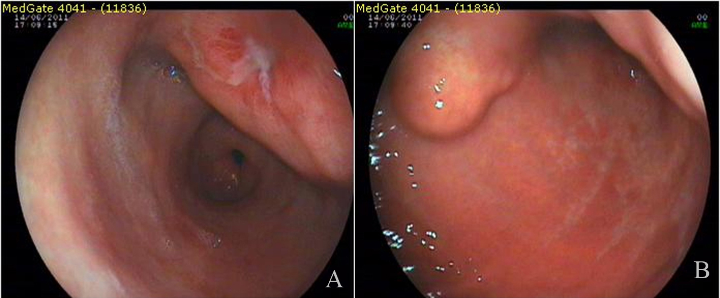
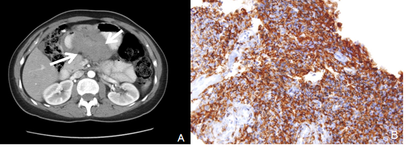
A 3 - 4 cm ulcerated polypoid mass in antrum and polypoid lesion with an impression of submucosal localization over the distal corpus (Fig. 1) were found in endoscopy in a female patient complaining of dyspeptic problems with an age of 54. Endoscopic biopsies revealed chronic inflammation and H.pylori was negative. First, abdominal tomography and then endoscopic ultrasonography were performed. Abdominal tomography showed a polypoid mass in the antrum with a diameter of 6 cm (Fig. 2A). Endoscopic ultrasonography yielded 5 - 6 cm transmural mass with serosal infiltration and heterogenous echogenity. Fine-needle aspiration biopsies were performed two times with a nondiagnostic material. Trucut biopsy was performed after cytologic examination yielding sample rich in lymphoid tissue. Histologic examination was reported as extranodal marginal-zone B-cell lymphoma with CD20 antigen (+) (Fig. 2B). The patient was consulted to Oncology Service for chemotherapy.
 Click for large image | Figure 1. Endoscopic appearance of the lesions. |
 Click for large image | Figure 2. A: Computerized tomography revealed antral mass; B: CD 20 positivity by immunohistochemical study in lymphoma cells. |
We can detect different endoscopic lesions in gastric lymphomas. Diagnostic value of fine needle aspiration biopsy in lymphoid tumors is limited.
Conflict of Interest
We have no conflict of interest disclosure.
Abbreviations
H.pylori: Helicobacter pylori
| References | ▴Top |
- Mehra M, Agarwal B. Endoscopic diagnosis and staging of mucosa-associated lymphoid tissue lymphoma. Curr Opin Gastroenterol. 2008;24(5):623-626.
pubmed doi - Ruskone-Fourmestraux A, Fischbach W, Aleman BM, Boot H, Du MQ, Megraud F, Montalban C,
et al . EGILS consensus report. Gastric extranodal marginal zone B-cell lymphoma of MALT. Gut. 2011;60(6):747-758.
pubmed doi - Zucca E, Bertoni F, Roggero E, Cavalli F. The gastric marginal zone B-cell lymphoma of MALT type. Blood. 2000;96(2):410-419.
pubmed
This is an open-access article distributed under the terms of the Creative Commons Attribution License, which permits unrestricted use, distribution, and reproduction in any medium, provided the original work is properly cited.
World Journal of Oncology is published by Elmer Press Inc.


